
On this page
If your textbook's replication fork shows one smooth new strand and one in pieces, the cause is one chemical rule about how DNA polymerase builds a chain.
What are Okazaki fragments?
Okazaki fragments are short stretches of new DNA made on the lagging strand and later joined into one continuous strand. Reiji Okazaki, Tsuneko Okazaki and colleagues reported evidence that new DNA chains are made discontinuously in a February 1968 paper in PNAS. We tell their story in Reiji and Tsuneko Okazaki and the fragments of DNA replication.
Their size depends on the organism. Prokaryotic Okazaki fragments are about 1,200 nucleotides long, while eukaryotic fragments are much shorter, with lengths set by the spacing of nucleosomes, according to a 2013 review by Lata Balakrishnan and Robert Bambara.
Why does DNA polymerase work only 5′ to 3′?
DNA polymerase can add nucleotides only to the free 3′-OH end of a growing strand, so every new strand grows in the 5′ to 3′ direction (OpenStax Biology 2e). Each incoming nucleotide arrives as a deoxyribonucleoside triphosphate; the enzyme cleaves off two of its phosphates and uses that energy to join the remaining 5′ phosphate to the 3′-OH of the chain (Penn State Biology 230 notes).
So the energy for each step travels in with the new nucleotide, not on the chain. That matters for proofreading: the same notes describe a second active site, a 3′ to 5′ exonuclease, that cuts a nucleotide off the 3′ end. After a mistake is clipped, the chain still ends in an ordinary 3′-OH and the next nucleotide brings its own energy, so copying carries on.
A chain growing 3′ to 5′ would instead carry the energy-rich triphosphate on its own end, and clipping a mistake would throw that energy away. That is one way to see why the one-direction rule makes sense.
Why is the lagging strand discontinuous?
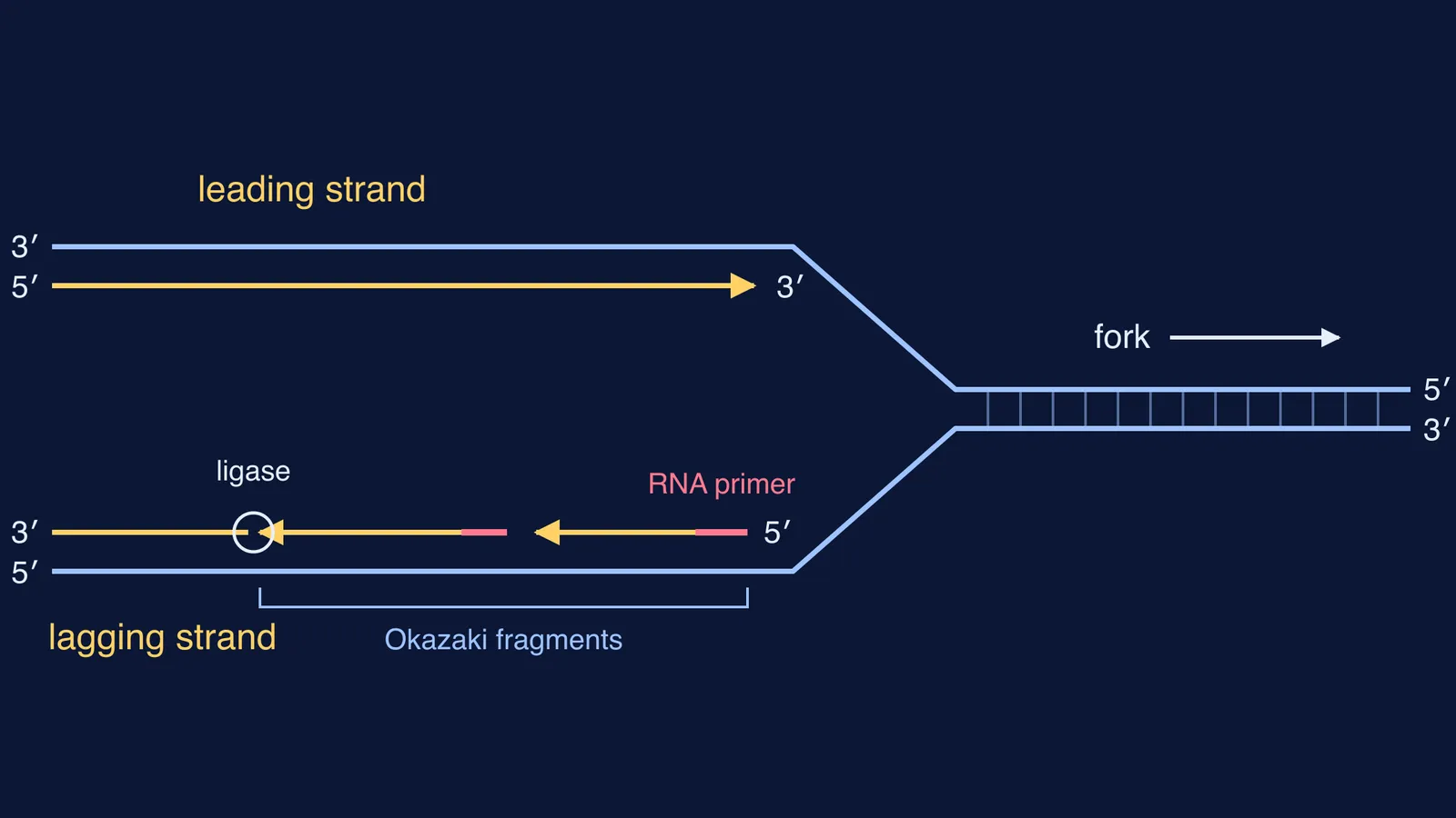
The two strands of the double helix are antiparallel: one runs 5′ to 3′, its partner 3′ to 5′ (more in what antiparallel means in DNA, and why 5′ to 3′ matters). At a replication fork both strands are unwound and copied at the same time, but the fork opens in only one direction.
On one template, 5′ to 3′ growth points the same way the fork moves. Polymerase follows the fork and makes one long, continuous leading strand.
On the other template, 5′ to 3′ growth points away from the fork. Polymerase makes a short piece, then has to start again nearer the fork as more template opens up. This is the lagging strand.
According to OpenStax Biology 2e, each fragment begins with an RNA primer, about five to ten nucleotides long, made by the enzyme primase.
What does DNA ligase do in replication?
In E. coli, the exonuclease activity of DNA polymerase I removes the RNA primers and the gaps are filled with DNA; DNA ligase then forms the phosphodiester bond between the 3′-OH end of one fragment and the 5′ phosphate end of the next (OpenStax Biology 2e). Without ligase, the lagging strand would stay a row of separate pieces with nicks between them.
The Balakrishnan and Bambara review describes the same basic scheme in prokaryotes and eukaryotes: the primer is pushed into a flap, the flap is removed, and the ends are ligated. Eukaryotes use many more enzymes, possibly in three pathways. In answers, keep the jobs apart: polymerase adds nucleotides; ligase only seals the backbone.
How do you label leading and lagging strands in an exam diagram?
Use these five steps whenever a question gives you a fork with strand ends marked.
Find which way the fork is moving: towards the still-closed parent duplex.
Write each new strand's ends, antiparallel to its template: a 3′ to 5′ template gets a 5′ to 3′ new strand.
If the new strand's 3′ end points toward the fork, it is the leading strand.
If its 3′ end points away from the fork, it is the lagging strand. Draw short arrows, each with a primer at its 5′ end, the newest piece nearest the fork.
Mark ligase at the nicks between finished pieces.
Try it on the diagram at the top. The fork moves right. The top template runs 3′ to 5′ left to right, so its new strand grows toward the fork: leading. The bottom template runs 5′ to 3′, so its new strand grows away from the fork, in pieces: lagging.
A quick number check: OpenStax Biology 2e gives about 1,000 nucleotides added per second in prokaryotes, and the Balakrishnan and Bambara review puts prokaryotic fragments at about 1,200 nucleotides. So a bacterial lagging-strand polymerase finishes a fragment in roughly 1.2 seconds.
Where to go next
If antiparallel strands still feel shaky, the Fermi Concept Library has a lesson for every concept in biology, so you can step back to DNA structure first. For drawing practice, follow how to draw the replication fork diagram step by step, then test yourself in the Fermi Problem Library, where a step-by-step solver explains the reasoning rather than printing an answer.