

On this page
Drawing the replication fork diagram is a staple of Class 12 biology board exams and a core concept for NEET. Examiners look for a few specific details: the correct Y-shape, the antiparallel polarity of the template strands, and the distinct ways the leading and lagging strands are built.
Before drawing, make sure you know the difference between nucleosides vs nucleotides: the chemistry of DNA and RNA units. You might also want to practice Chargaff's rule numericals to understand base pairing.
How to draw the replication fork diagram?
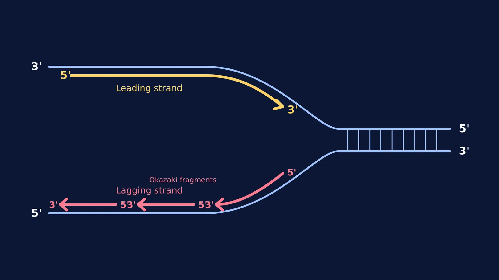
Draw the template: Start with a large Y-shape. The stem of the Y is the unbroken parental double helix, and the two open arms are the unwound template strands.
Label the polarity: This is where most students lose marks. Label one template strand as 3′ at its unwound end and 5′ at the stem. Label the other template strand as 5′ at the unwound end and 3′ at the stem.
Draw the leading strand: On the template strand that runs 3′ to 5′, draw a single, unbroken arrow pointing *into* the fork. Label its tail as 5′ and its arrowhead as 3′. This shows continuous synthesis.
Draw the lagging strand: On the template strand that runs 5′ to 3′, draw two or three short arrows pointing *away* from the fork. Label each fragment with 5′ at the tail and 3′ at the arrowhead. This shows discontinuous synthesis.
Which enzymes work at the replication fork?
A whole team of enzymes operates at the replication fork to keep the process moving. First, DNA helicase unzips the parental double helix. According to *Molecular Biology of the Cell*, these helicases can pry apart the DNA at rates of up to 1000 nucleotide pairs per second.
Once the strands are separated, they must be primed before the new DNA can be built. DNA polymerase cannot start a new chain from scratch; it requires an existing 3′ end to add onto. A special enzyme called primase builds a short RNA primer to provide this starting point. On the leading strand, this only needs to happen once. On the lagging strand, primase must constantly lay down new primers for each Okazaki fragment. Finally, an enzyme called DNA ligase seals the gaps between the short Okazaki fragments, joining them together into one continuous DNA molecule.
Why is one strand continuous and the other discontinuous?
DNA polymerase, the enzyme that builds the new strand, is a one-way machine. It can only add nucleotides to the 3′ end of a growing chain. Because the two parental template strands run in opposite directions (they are antiparallel), the enzyme must work differently on each side.
The leading strand grows smoothly towards the unwinding fork because it is moving in the correct 5′ to 3′ direction. However, the lagging strand must grow away from the fork. As the fork opens further, the enzyme must jump back to the newly exposed template and start a new fragment.
If you are confused by how the enzymes work together, Fermi's AI tutor teaches rather than answers. It holds a line of reasoning across a whole session, notices where you are stuck, and draws the figure to help you see it.
What is the speed of DNA replication?
The enzymes at the replication fork work at astonishing speeds. According to *Molecular Biology of the Cell*, the bacterial replication machine can duplicate DNA at rates up to 1000 nucleotides per second. Eukaryotic replication forks, such as those in human cells, move about one-tenth as fast, likely because they have to navigate around nucleosomes.
The short segments of DNA on the lagging strand are called Okazaki fragments. If you are curious about the researchers who discovered them, read about Reiji and Tsuneko Okazaki and the fragments of DNA replication. In bacteria, these fragments are 1000 to 2000 nucleotides long, but in eukaryotes, they are much shorter, typically only 100 to 200 nucleotides long.
A checklist for your exam diagram
Before you hand in your paper, run your diagram through this quick checklist:
[ ] Are your template strands antiparallel (one 3′→5′, the other 5′→3′)?
[ ] Does every new DNA arrow point from 5′ to 3′?
[ ] Is the continuous strand growing towards the fork?
[ ] Are the Okazaki fragments growing away from the fork?
[ ] Have you added the standard caption: "Replicating Fork showing continuous and discontinuous synthesis"?
You can review the specific roles of helicase and ligase in the Fermi Concept Library, where you will find every concept in biology with its own lesson and its place in the map.


