On this page
Why is octopus blood blue instead of red?
When you cut your finger, blood flows crimson red because red blood cells are packed with iron-containing haemoglobin. In an octopus, cuttlefish, or squid, blood is transparent in oxygen-depleted tissues and turns vivid cyan blue the moment it absorbs oxygen at the gills.
In 1878, Belgian physiologist Léon Fredericq studied the common octopus (*Octopus vulgaris*) at Brittany's Roscoff Biological Station, as recounted in Experientia. Fredericq proved that the respiratory pigment used copper rather than iron, coining the name "haemocyanin" from Greek *haima* (blood) and *kyanos* (blue). Haemocyanin is now recognized as Earth's second most common respiratory pigment, found throughout molluscs and arthropods.
How does haemocyanin bind oxygen at the molecular level?
Haemocyanin differs fundamentally from haemoglobin. In the Journal of Molecular Biology, Marianne E. Cuff and colleagues resolved the crystal structure of an octopus haemocyanin functional unit. Each oxygen-binding site consists of a binuclear copper centre coordinated directly by six histidine residues—three per copper atom—with no porphyrin ring.
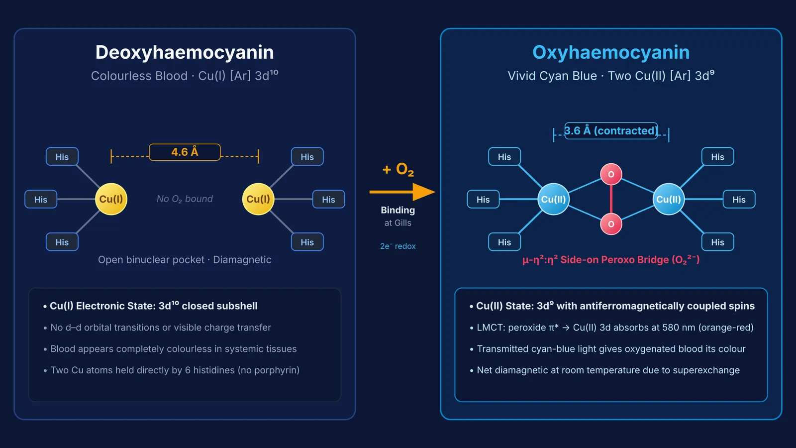
In the deoxygenated state, both copper atoms are Cu(I) with a full d-subshell ([Ar] 3d¹⁰). Lacking unpaired d-electrons, Cu(I) cannot undergo d–d transitions, leaving deoxygenated blood completely colourless.
When oxygen binds, it coordinates in a side-on bridging geometry (μ-η²:η² peroxo bridge) between the coppers. As detailed in Chemical Reviews by Felix Tuczek and Edward I. Solomon, a two-electron redox transfer oxidises the coppers to Cu(II) (3d⁹) and reduces oxygen to peroxide (O₂²⁻), contracting the Cu–Cu distance from 4.6 Å to 3.6 Å.
This configuration creates an intense ligand-to-metal charge transfer (LMCT) from peroxide π* orbitals to copper(II) 3d orbitals. This transition absorbs strongly near 580 nm (orange-red) and 345 nm (near-ultraviolet), transmitting cyan-blue light. Strong antiferromagnetic coupling between the Cu(II) centres makes oxyhaemocyanin diamagnetic.
How does haemocyanin compare with human haemoglobin?
Key structural and functional differences include:
Active metal ion: Haemoglobin uses iron (Fe(II)), whereas haemocyanin uses copper (Cu(I) in deoxy, Cu(II) in oxy).
Chemical scaffold: Haemoglobin binds iron inside a protoporphyrin IX ring, whereas haemocyanin coordinates copper directly to histidine side chains.
Binding stoichiometry: Haemoglobin binds one O₂ per iron atom (1:1 ratio), while haemocyanin requires two copper atoms per O₂ molecule (2:1 ratio).
Quaternary architecture: Human haemoglobin is an intracellular 64.5 kDa tetramer. Cephalopod haemocyanin is a massive extracellular multimer of roughly 3.5 to 3.8 MDa dissolved in hemolymph, as reviewed in Advances in Protein Chemistry60545-8).
Oxygen carrying capacity: Human blood carries roughly 20 mL of O₂ per 100 mL of blood, whereas octopus hemolymph carries only about 3 to 5 mL of O₂ per 100 mL.
To see how vertebrates tailor oxygen delivery using intracellular pigments across different tissues, compare this mechanism with our guide on haemoglobin vs myoglobin.
Why did cephalopods keep copper blood in freezing oceans?
In freezing polar waters, haemocyanin remains remarkably functional. A 2015 study in Frontiers in Zoology led by Michael Oellermann showed that the Antarctic octopus *Pareledone charcoti* maintains roughly 40% higher blood haemocyanin concentrations than temperate octopuses to sustain oxygen delivery at temperatures below 0 °C.
Because hemolymph carries limited oxygen, octopuses rely on a closed circulatory system powered by three hearts: two branchial hearts pushing blood through gills and one systemic heart driving it through the body. For comparative mechanics, read our analysis of open vs closed circulatory systems.
Cephalopod haemocyanin also exhibits a steep Bohr shift, releasing oxygen rapidly in active muscle as pH falls, a mechanism explored in our review of the Bohr effect.
How to solve respiratory pigment questions in competitive exams?
Bioinorganic exam questions frequently test coordination chemistry alongside animal physiology.
Use this 4-step checklist when answering bioinorganic questions:
Identify metal oxidation states: Fe(II) in haemoglobin; Cu(I) in deoxyhaemocyanin and Cu(II) in oxyhaemocyanin.
Check coordination geometry: Planar porphyrin ring in haemoglobin versus six histidine ligands in haemocyanin.
Verify stoichiometry: 1:1 metal-to-dioxygen ratio in haemoglobin versus 2:1 in haemocyanin.
Determine magnetic states: Deoxyhaemocyanin is colourless and diamagnetic (3d¹⁰); oxyhaemocyanin is blue and diamagnetic due to antiferromagnetic coupling.
When reviewing biochemical pathways and coordination complexes, the Fermi Concept Library maps every concept across biology and chemistry with dedicated lessons. For exam practice, the Fermi Problem Library offers worked problems with a step-by-step solver that explains the reasoning rather than printing an answer.