On this page
Peel a ripe tomato and the flesh almost falls apart in your fingers. Bite a raw carrot and it holds firm. Part of that difference lies in a thin layer you meet in Class 11 biology: the middle lamella, the shared layer that sticks one plant cell to the next.
What is the middle lamella made of?
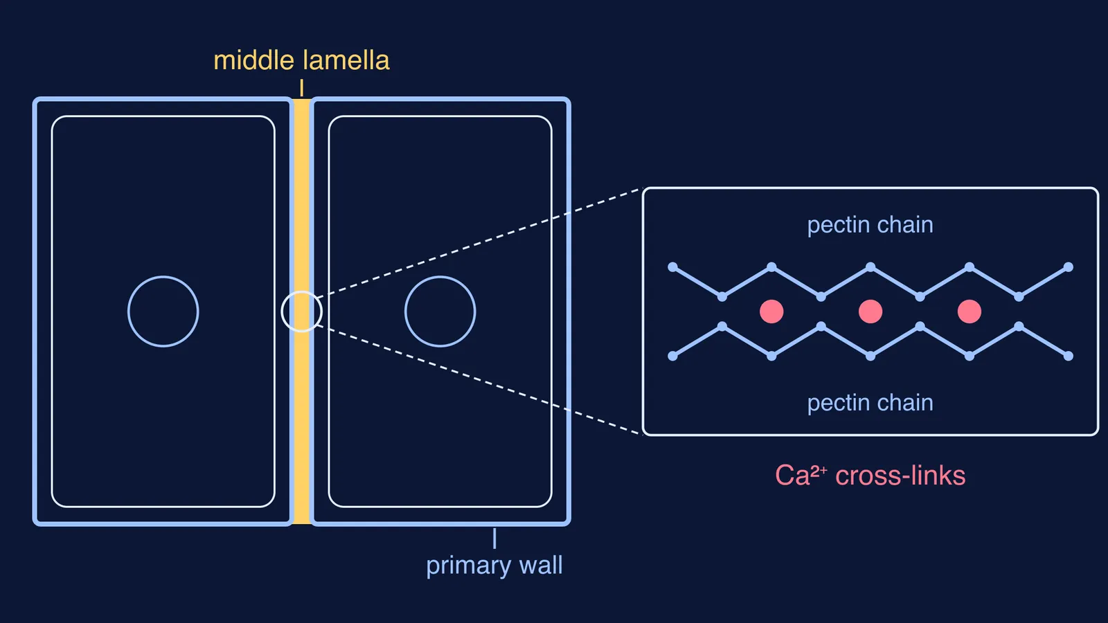
The short answer is pectin held together by calcium. The Paul Knox cell wall lab at the University of Leeds describes the layer between plant cells as an intercellular matrix that is "thought to consist largely of pectic polymers".
The key pectin is homogalacturonan, a chain of linked galacturonic acid units that can carry methyl groups on their carboxyl groups, as set out in the background to a 2014 Plant Physiology study by Domozych and colleagues, which looks at pectin in a green alga. Where a stretch of the chain has no methyl groups, the same paper explains, calcium ions bridge neighbouring chains in so-called egg-box complexes and the pectin sets into a gel.
That calcium-pectin gel is what your textbook shortens to "calcium pectate". The same 2014 paper reports that pectin with few or no methyl groups is most abundant in the middle lamella, especially where cells meet and at the corners of the air spaces between them.
Pectin is a big part of the wall in general, not just the glue layer. In Arabidopsis leaves, roughly 50% of the cell wall is pectin, according to a 2015 review by Bou Daher and Braybrook in Frontiers in Plant Science.
How does the cell plate become the middle lamella?
A plant cell cannot pinch in two, so it builds a new partition across its middle. For the plant-versus-animal contrast, see our post on how cytokinesis differs in plant and animal cells.
Here is the sequence, using the Bou Daher and Braybrook review and the Leeds cell wall lab:
During cell division, a large, directed stream of vesicles carrying homogalacturonan pectin is delivered to the middle of the cell.
The vesicles fuse into a partition called the cell plate, built across the middle of the cell.
Cellulose synthases then arrive and hemicellulose delivery begins, so each daughter cell builds its own new primary wall.
The cell plate between them becomes the pectin-rich middle lamella, shared by both cells.
An enzyme called pectin methylesterase removes methyl groups from the pectin. The stripped chains are cross-linked by calcium, and the layer firms up.
One honest detail: the Domozych paper notes that the material laid down at the cell plate would not be enough on its own to keep up with the large increase in wall area as the cell expands. The cell plate is the start of the middle lamella, not all of it.
Why do ripe fruits go soft?
The glue can also be undone. Enzymes called polygalacturonases break down the pectin backbone, and the 2015 review links them to fruit ripening, leaf and fruit drop (abscission) and the splitting open of seed pods.
Calcium matters just as much. In ripe tomato pericarp, calcium-linked pectin is a major adhesive bond, and soaking the tissue in a chemical that grabs calcium can quickly make the cells separate completely, the Leeds lab notes. That is the tomato in your fingers.
Where do students lose marks on the middle lamella?
Check your answer against this list:
Order of layers. From the outside of a mature cell inwards: middle lamella, primary wall, secondary wall (only in some cells), then the plasma membrane. The middle lamella is the outermost layer, as the overview on Wikipedia summarises from standard textbooks.
Chemistry. Pectin and calcium, not cellulose. Cellulose belongs to the primary and secondary walls; our post on what the cell wall is made of in plants, fungi and bacteria lays out the full comparison.
Ownership. The middle lamella is shared by two cells. It is not part of one cell's wall.
Origin. It comes from the cell plate formed during cytokinesis, not from the old parent wall.
Links between cells. Do not confuse the glue with the channels; plasmodesmata connect neighbouring plant cells for a different job.
A model three-mark answer you can adapt:
"The middle lamella is the layer that holds adjacent plant cells together. It is made mainly of pectin cross-linked by calcium ions (calcium pectate). It forms from the cell plate laid down between daughter cells during cytokinesis, and each daughter cell then deposits its own primary wall on either side of it."
Try this before your next test: draw two cells side by side from memory, label all four layers in order, and write one line on where each came from. If you get stuck on any layer, the Fermi Concept Library covers every concept in biology, each with its own lesson and its place in the map.
For mixed questions on layers and division stages, the Fermi Problem Library has a step-by-step solver that explains the reasoning rather than printing an answer.