On this page
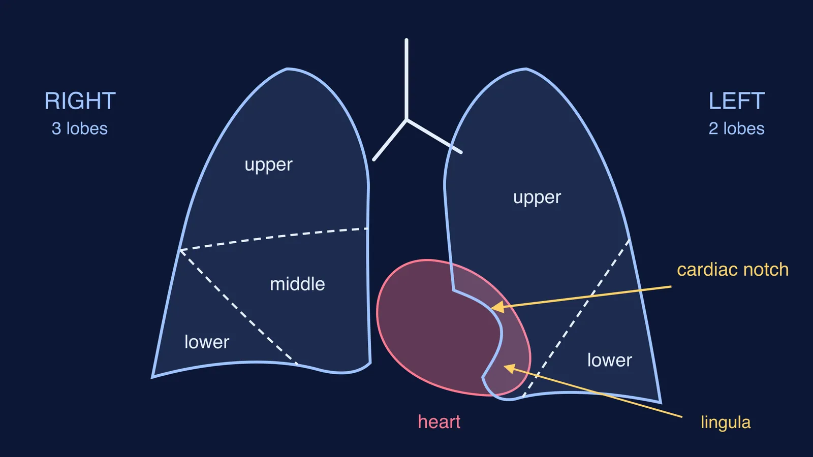
Your two lungs are not mirror images. The right one has three lobes, the left one has two, and the left one carries a dent called the cardiac notch where the heart sits. If you have ever drawn them as identical twins in a practical record, this is the detail to fix first.
Why is the left lung smaller than the right lung?
The left lung shares space in the chest with the heart, as the Wikipedia article on the lung puts it, so it has less room on its side. The open textbook OpenStax Anatomy and Physiology says it plainly: the right lung is shorter and wider than the left, and the left lung occupies a smaller volume than the right.
The right lung accounts for about 55% of total lung volume and function, according to IMAIOS e-Anatomy. The same reference explains why the right lung is shorter: the right half of the diaphragm sits higher than the left, pushed up by the liver beneath it.
What is the cardiac notch of the left lung?
The cardiac notch is an indentation on the surface of the left lung that makes space for the heart, as defined in OpenStax Anatomy and Physiology.
It sits on the front (anterior) border of the left lung, where the apex of the heart presses in, according to TeachMeAnatomy. On the inner surface there is also a hollow where the heart sits, the cardiac impression, which is deeper and larger on the left lung than on the right, as the Wikipedia article on the lung notes.
Just below the notch, the upper lobe of the left lung sends out a tongue-shaped projection called the lingula, as described by the Manipal anatomy museum. The Wikipedia entry on the lingula describes it as a projection of the upper lobe that is an anatomical parallel to the right lung's middle lobe. So it is part of the upper lobe, not a third lobe.
How many lobes and fissures does each lung have?
Fissures are the deep grooves that divide a lung into lobes. Here is the comparison, from OpenStax and the Wikipedia lung article:
Right lung: three lobes (superior, middle, inferior) and two fissures (oblique and horizontal).
Left lung: two lobes (superior, inferior) and one fissure (oblique), plus the cardiac notch and the lingula.
One more asymmetry sits in the airway. The right main bronchus is wider, shorter and more vertical than the left, which is why inhaled foreign objects more often end up in the right lung, according to IMAIOS e-Anatomy. Learn the lobes and this bronchus fact together.
For the bigger picture, see why your heart runs two loops in double circulation and breathing vs cellular respiration.
How do you draw and label the lungs in an exam?
Diagrams are usually drawn in front view, as if the person is facing you. That means the person's right lung appears on the left side of your page. It is easy to mix the two up, so decide this before you start.
A drawing checklist:
Draw the trachea and split it into two bronchi, the right one (page left) shorter and steeper.
Draw the right lung (page left) broad and a little shorter at the base.
Draw the left lung (page right) narrower, with a scoop out of its inner front edge: the cardiac notch.
Sketch the heart in the notch, tilted towards page right.
On the right lung, draw the horizontal and oblique fissures and label upper, middle and lower lobes.
On the left lung, draw only the oblique fissure, label upper and lower lobes, and mark the lingula below the notch.
Write "right" and "left" for the person, not the page, and check them last.
Test yourself: true or false?
The left lung has a horizontal fissure. False: only the oblique.
The lingula is a third lobe. False: it belongs to the upper lobe.
The cardiac notch is on the right lung. False: it is on the left.
If the shape still will not settle, the Fermi Concept Library has a lesson for every concept in biology, each with its place in the map, so you can see what the lungs connect to. Fermi's AI tutor can also draw the figure with you and check your understanding as it goes; our guide to studying biology diagrams with an AI tutor shows how to use that well.