On this page
If a biology question asks what holds a chromosome to the spindle, the answer is not the centromere itself. It is a small structure built on top of it. Once you see its job, metaphase and anaphase stop being lists to memorise and start making sense.
What is a kinetochore?
The NCERT Class 11 biology chapter on the cell cycle defines kinetochores as "small disc-shaped structures at the surface of the centromeres" that "serve as the sites of attachment of spindle fibres to the chromosomes" (NCERT Biology Class 11, Chapter 10).
Researchers describe the same thing at the molecular level. Kinetochores are large protein assemblies that connect chromosomes to the microtubules of the spindle, so that the copied genome is shared between two daughter cells (Musacchio and Desai, Biology, 2017). An earlier review reported that studies had identified more than 80 kinetochore components (Cheeseman and Desai, Nature Reviews Molecular Cell Biology, 2008). The disc in your textbook is a simplified picture of a busy protein machine.
Kinetochore vs centromere: what is the difference?
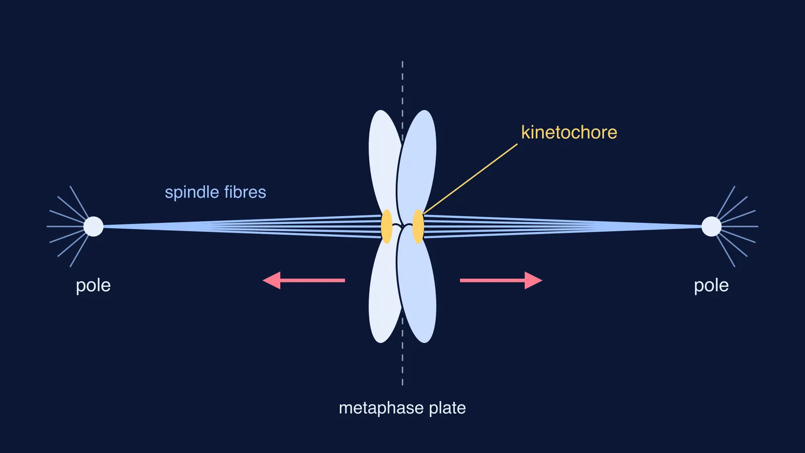
The centromere is the region where the two sister chromatids of a chromosome are held together (NCERT, Chapter 10). The kinetochore is the structure on the surface of that region. Spindle fibres attach to the kinetochore.
At metaphase, each chromosome has two kinetochores: one on each sister chromatid, facing opposite poles. Keep that detail in mind; everything that follows depends on it. If you mix up sister chromatids and homologous chromosomes, our post on sister chromatids vs homologous chromosomes sorts out which pair splits when.
What does the kinetochore do in each stage of mitosis?
Follow one chromosome through mitosis. The stage descriptions below come from NCERT Chapter 10; the force and checkpoint details come from Musacchio and Desai.
Prophase into metaphase: the nuclear envelope breaks down, and spindle fibres from the two poles attach to the kinetochores.
Metaphase: one chromatid is connected by its kinetochore to fibres from one pole, and its sister to fibres from the opposite pole. The chromosomes line up at the metaphase plate.
The check before splitting: the spindle assembly checkpoint stops the cell leaving mitosis while any kinetochore is unattached or wrongly attached.
Anaphase: each chromosome splits and the daughter chromatids move to opposite poles. The centromere stays at the leading edge, facing the pole, and the arms trail behind.
How does a kinetochore pull? It holds on to the end of a microtubule as it shrinks. A depolymerizing microtubule generates enough force to move a chromosome bound through its kinetochore (Musacchio and Desai). The fibre gets shorter, and the chromatid is dragged towards the pole.
How does a cell catch a wrong attachment?
Sometimes both sister kinetochores grab fibres from the same pole. Kinetochores control feedback mechanisms that correct wrong microtubule attachments (Musacchio and Desai). The same review says tension, within the kinetochore or between sister kinetochores, helps the cell tell correct attachments from wrong ones. In the simple picture, a pair pulled from both sides is under tension; a pair pulled from one side is not. Meanwhile the checkpoint holds anaphase back.
Your textbook does not go into the molecular details. But the idea explains why metaphase looks so tidy: the cell does not move on while any kinetochore is unattached or wrongly attached.
How to answer kinetochore questions in exams
Here is the kind of reasoning a counting question needs.
Question: A cell at metaphase has 8 chromosomes. How many chromatids and kinetochores are there?
Reasoning: Each metaphase chromosome has two sister chromatids, and each chromatid has its own kinetochore. So 8 chromosomes give 16 chromatids and 16 kinetochores. At anaphase those 16 chromatids become daughter chromosomes, 8 moving to each pole.
Before a test, check that you can:
state the NCERT definition in one sentence, using "disc-shaped", "centromere" and "spindle fibres"
explain why a kinetochore is not the same as a centromere
draw metaphase with a kinetochore on the outer face of each chromatid and fibres from opposite poles
draw anaphase with the centromere leading and the arms trailing
If you are unsure which stage a diagram shows, read our guide on how to identify mitosis and meiosis stages from a diagram. For why chromosomes are studied at this stage, see why metaphase is the best stage to study chromosome morphology.
If the chain from centromere to fibre to pole still feels shaky, the Fermi Concept Library gives every biology concept its own lesson and its place in the map. To test yourself, the Fermi Problem Library has worked problems with a step-by-step solver that explains the reasoning rather than printing an answer.
Next time you draw metaphase, put the two kinetochores in first, then the fibres. The rest of the diagram follows from them.