

On this page
When a cell prepares to divide, it must move its DNA safely without breaking it. A single human DNA molecule stretched out would measure several centimetres. During metaphase, this same strand is packed into a dense package just 2 to 10 micrometres (µm) long (source). This remarkable 10,000-fold compaction means the chromosomes are at their shortest and thickest, making their physical shape—their morphology—clearly visible under a standard light microscope.
For example, human Chromosome 1, the largest, measures around 10 µm in metaphase, while the smallest, such as Chromosomes 21 and 22, are just 2 µm long.
How are chromosomes classified by shape?
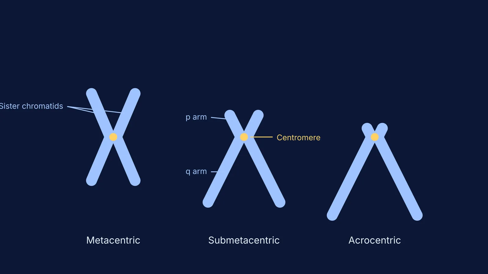
At this stage, the two sister chromatids are perfectly defined and joined at the centromere. Because the sister chromatids are held tightly together, the arms splay outward, forming the classic "X" shape we associate with chromosomes. However, not all chromosomes look identical. Depending on where the centromere is positioned, the arms can be equal in length (metacentric), slightly unequal (submetacentric), or extremely skewed with almost no "p" (short) arm at all (acrocentric). This distinct morphology allows scientists to arrange them into an ordered karyogram, sorted from the largest to the smallest.
See **Chromosome types by centromere position: metacentric to telocentric** for more on how these shapes are classified.
How do chromosomes align at the metaphase plate?
In earlier stages like prophase, chromosomes look like a tangled ball of threads. In metaphase, the nuclear envelope has completely broken down, and microtubules from the spindle poles attach to the chromosomes' kinetochores (Wikipedia). The chromosomes line up in a single plane across the centre of the cell, known as the metaphase plate or equatorial plate.
The metaphase plate is not a physical structure, but an imaginary equator equidistant from the two spindle poles. As the microtubules tug on the kinetochores—protein structures built on the centromeres—the chromosomes are pulled into this central alignment. This delicate balance of tension ensures that when the cell finally transitions into anaphase, each daughter cell will receive exactly one copy of every chromatid.
This orderly line-up is crucial for biological study. Because the chromosomes are spread out in a single layer rather than clumped together, they do not overlap. This makes it straightforward for geneticists to count them accurately and spot large-scale structural changes, such as a missing section (deletion) or an extra copy of a whole chromosome, as seen in Down syndrome.
Read more about how the spindle fibres guide this process in **Cell cycle, mitosis and meiosis: stages and chromosome counts**.
How do labs pause cells in metaphase for karyotyping?
To study human chromosomes—a process called karyotyping—scientists take a blood sample and encourage the white blood cells to divide (Wikipedia). However, metaphase is just a brief window in the cell cycle. To get enough cells in this exact stage, labs use a chemical called colchicine.
Colchicine is an alkaloid extracted from the autumn crocus plant. It acts as a mitotic poison by preventing the polymerisation of tubulin into microtubules (source). Without the microtubule spindle fibres to attach to the kinetochores and pull the sister chromatids apart into anaphase, the cell's internal machinery stalls. The cell cycle effectively stops dead at metaphase.
Once the cells are arrested in this phase, the lab treats them with a dilute, hypotonic solution. Water rushes into the cells via osmosis, causing them to swell. This swelling physically spreads the condensed chromosomes out within the cell membrane. Finally, the cells are dropped onto a glass slide, bursting open and scattering the chromosomes so they lie flat. They are then stained with specific dyes—most commonly Giemsa stain—which produces a unique pattern of light and dark bands (G-bands) across each chromosome's arms. This banding pattern acts like a barcode, confirming the identity of each chromosome and highlighting any subtle genetic rearrangements.
If you are preparing for exams, understanding these practical techniques is just as important as the theory. The Fermi Concept Library maps out every concept in biology, letting you explore how chemicals like colchicine interact with the cell machinery. You can also read about how this looks under a microscope in **Onion root tip mitosis: the Class 12 practical slide explained**.


