On this page
In ICSE Class 10 Biology, the ear sits in the sense-organs chapter next to the eye. You need two things: the order in which sound travels, and a neat labelled diagram. Here are both, checked against the US National Institute on Deafness and Other Communication Disorders (NIDCD) and hospital guides. Syllabus wording can change, so confirm the current list with CISCE or your teacher.
What are the three parts of the human ear?
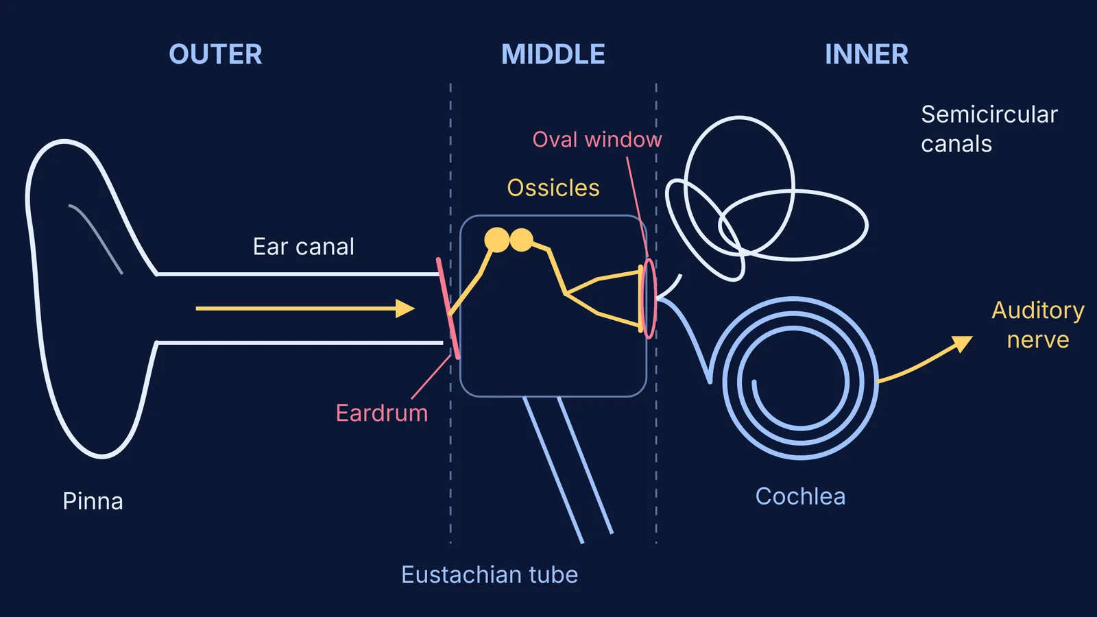
The ear is divided into the outer ear, the middle ear and the inner ear, and the eardrum is the landmark between the first two. Each definition below comes from the University Hospitals guide to ear anatomy.
Outer (external) ear: the pinna, the outside part of the ear that you can see, and the ear canal, the tube that connects the outer ear to the middle ear.
Middle ear: the space behind the eardrum. The tympanic membrane, or eardrum, divides the external ear from the middle ear, and the middle ear holds the three ossicles and the opening of the Eustachian tube.
Inner ear: the cochlea, which contains the nerves for hearing, plus the vestibule and semicircular canals, which contain receptors for balance.
How does sound travel from the pinna to the cochlea?
The NIDCD page How Do We Hear? sets out the sequence step by step. In exam language, it runs like this:
Pinna: sound waves enter the outer ear.
Ear canal: the waves travel down this narrow passage to the eardrum.
Eardrum (tympanum): it vibrates with the incoming sound waves.
Ossicles: the malleus, incus and stapes pick up the vibration; NIDCD says these middle-ear bones amplify, or increase, the sound vibrations.
Oval window: the stapes is attached to the membrane of the oval window, the opening between the middle ear and the inner ear (Wikipedia: Ossicles).
Cochlea: the fluid inside this snail-shaped structure ripples, a travelling wave runs along the basilar membrane, and as the hair cells riding that wave move, their stereocilia bend, which creates an electrical signal.
Auditory nerve: it carries the signal to the brain, which turns it into a sound we recognise.
Memory hook: air in the outer ear, bone in the middle, fluid in the inner ear.
What do the ossicles and the Eustachian tube do?
The three ossicles are the malleus, incus and stapes, names usually translated as the hammer, anvil and stirrup, and the stapes is the smallest bone in the body (Wikipedia: Ossicles). Through lever action and by spreading the force over a smaller area, the chain makes the vibrations reaching the inner ear stronger but smaller in movement (same source). For the shape of each bone, see our guide to the malleus, incus and stapes.
The Eustachian tube is a canal that links the middle ear with the back of the nose and helps to equalise the pressure in the middle ear (University Hospitals). By controlling the pressure behind the eardrum, it also helps keep the middle ear free of fluid (MedlinePlus).
How does the inner ear keep you balanced?
The balance part of the inner ear belongs to the labyrinth, a maze-like structure of bone and soft tissue (NIDCD: Balance Disorders). The semicircular canals contain three fluid-filled ducts that form loops roughly at right angles to one another, and they tell your brain when your head rotates (same source). Two fluid-filled pouches, the utricle and saccule, tell your brain the position of your head with respect to gravity, such as whether you are sitting up or lying down (same source).
To revise the ear alongside the eye and the rest of the sense organs, the Fermi Concept Library has every concept in physics, chemistry, biology, mathematics and computing, each with its own lesson and its place in the map.
Labelled diagram checklist for ICSE Class 10
Draw sound entering from the left and bracket the outer, middle and inner regions. Then tick off these labels, each with a one-line function:
Pinna: catches sound waves and leads them into the ear canal.
Ear canal: carries sound to the eardrum (its wax has a job too: read why we have ear wax).
Eardrum (tympanum): vibrates; separates the outer ear from the middle ear.
Malleus (hammer): attached to the eardrum.
Incus (anvil): links the malleus to the stapes.
Stapes (stirrup): smallest bone; attached to the oval window.
Oval window: the entry into the inner ear.
Eustachian tube: links the middle ear to the back of the nose; equalises pressure.
Cochlea: coiled and fluid-filled; hair cells turn vibrations into electrical signals.
Semicircular canals: three loops roughly at right angles; sense rotation of the head.
Vestibule (utricle and saccule): senses head position relative to gravity.
Auditory nerve: carries signals to the brain.
Keep label lines ruled and never crossing, with the names lined up on one side. Then try our hearing range tone test to find where your own hearing fades.