
On this page
If a question asks you to sort joints into immovable, slightly movable and freely movable, it is really asking one thing: what holds the two bones together? Change the tissue in the gap and you change how much the joint can move.
What are the three structural types of joints?
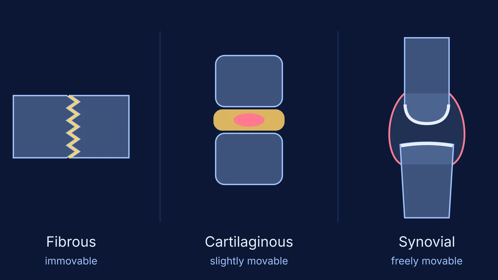
The NCERT Class 11 Biology chapter on locomotion and movement says joints "have been classified into three major structural forms, namely, fibrous, cartilaginous and synovial" (NCERT Biology Class 11, Chapter 17). Here is what sits between the bones in each one.
Fibrous joints: "A fibrous joint is where the adjacent bones are united by fibrous connective tissue" (OpenStax Anatomy and Physiology 2e). In the skull, the flat bones "fuse end-to-end with the help of dense fibrous connective tissues in the form of sutures" (NCERT).
Cartilaginous joints: "At a cartilaginous joint, the bones are joined by hyaline cartilage or fibrocartilage" (OpenStax). NCERT's example is the joint between adjacent vertebrae.
Synovial joints: the bones are not directly connected. Instead they meet "within a joint cavity that is filled with a lubricating fluid" (OpenStax). NCERT names the knee as a hinge joint of this type.
The pattern to remember: fibrous tissue holds bones tight, a pad of cartilage gives a little, and a fluid-filled gap lets the bones slide.
Which joints are immovable, slightly movable and freely movable?
OpenStax calls an immobile or nearly immobile joint a synarthrosis, a joint with limited mobility an amphiarthrosis, and a freely mobile joint a diarthrosis (OpenStax, classification of joints).
Immovable (synarthrosis): skull sutures. NCERT states that fibrous joints "do not allow any movement" (NCERT). "A suture is the narrow fibrous joint found between most bones of the skull" (OpenStax, fibrous joints).
Slightly movable (amphiarthrosis): joints between vertebrae. NCERT says the joint between adjacent vertebrae "permits limited movements" (NCERT). There, "a thick pad of fibrocartilage called an intervertebral disc strongly unites the adjacent vertebrae" (OpenStax, cartilaginous joints).
Freely movable (diarthrosis): the knee, shoulder and others. A synovial joint's fluid-filled cavity "allows considerable movement" (NCERT).
For the skull side of this, see our guide to the cranial and facial bones of the human skull.
What structure makes a synovial joint move freely?
"Synovial joints are the most common type of joint in the body" (OpenStax, synovial joints). Four parts do the work:
Articular cartilage: a thin layer of hyaline cartilage covering the end of each bone.
Articular capsule: a sleeve of fibrous connective tissue attached to both bones, enclosing the cavity.
Synovial membrane: a thin lining on the inside of the capsule.
Synovial fluid: a thick, slimy fluid inside the cavity that provides lubrication.
Ligaments, strong bands of fibrous connective tissue, anchor the bones together and support the joint. If you mix up ligaments and tendons, read tendon vs ligament and what each connects.
Learn the NCERT list of synovial joints as pairs: ball and socket (humerus and pectoral girdle), hinge (knee), pivot (atlas and axis), gliding (between the carpals) and saddle (carpal and metacarpal of the thumb) (NCERT). We test each with a movement check in ball and socket, hinge and pivot joints explained.
How do you classify a joint you have not seen before?
Use this three-step test.
Is there a fluid-filled cavity between the bones? If yes, it is synovial and freely movable. Stop.
If no, is the gap filled with cartilage (hyaline or fibrocartilage)? If yes, it is cartilaginous and usually slightly movable.
If no, the bones are held by fibrous connective tissue: fibrous, and immovable or nearly so.
Worked examples that catch students out:
Pubic symphysis: NCERT says the two halves of the pelvic girdle meet "to form the pubic symphysis containing fibrous cartilage" (NCERT). Cartilage and no cavity, so cartilaginous and slightly movable.
A tooth in its socket: OpenStax calls this a gomphosis, "the narrow fibrous joint between the roots of a tooth and the bony socket" (OpenStax). Fibrous.
A baby's soft spots: fontanelles are broad areas of connective tissue between skull bones that "greatly decrease in width during the first year after birth" (OpenStax). Still fibrous.
A growth plate: OpenStax describes the epiphyseal plate of a growing long bone as "a temporary synchondrosis", a cartilaginous joint later replaced by bone. OpenStax classes it as immobile, so cartilage does not always mean movement (OpenStax).
If fibrocartilage and hyaline cartilage still feel vague, the Fermi Concept Library gives every biology concept its own lesson and shows its place in the map. To practise, the Fermi Problem Library has worked problems with a step-by-step solver that explains the reasoning rather than printing an answer.
Your next step: pick five joints from your textbook diagrams, run each through the three-step test, and name the tissue out loud before you name the type.