

On this page
The human skull is a complex and vital structure that protects the brain and supports the face. It is the crowning component of the axial skeleton (see our guide to The human skeleton: counting axial and appendicular bones for the full 206-bone breakdown). But how many bones make up the adult human skull?
The answer is 22 bones, which anatomists divide into two main groups: the cranial bones and the facial bones 1. (Note that this count excludes the six tiny auditory ossicles in the middle ear and the hyoid bone in the neck).
Here is a full breakdown of the cranial and facial bones, the unique role of the mandible, and the fibrous joints that hold the skull together.
How many bones in the human skull cranial facial?
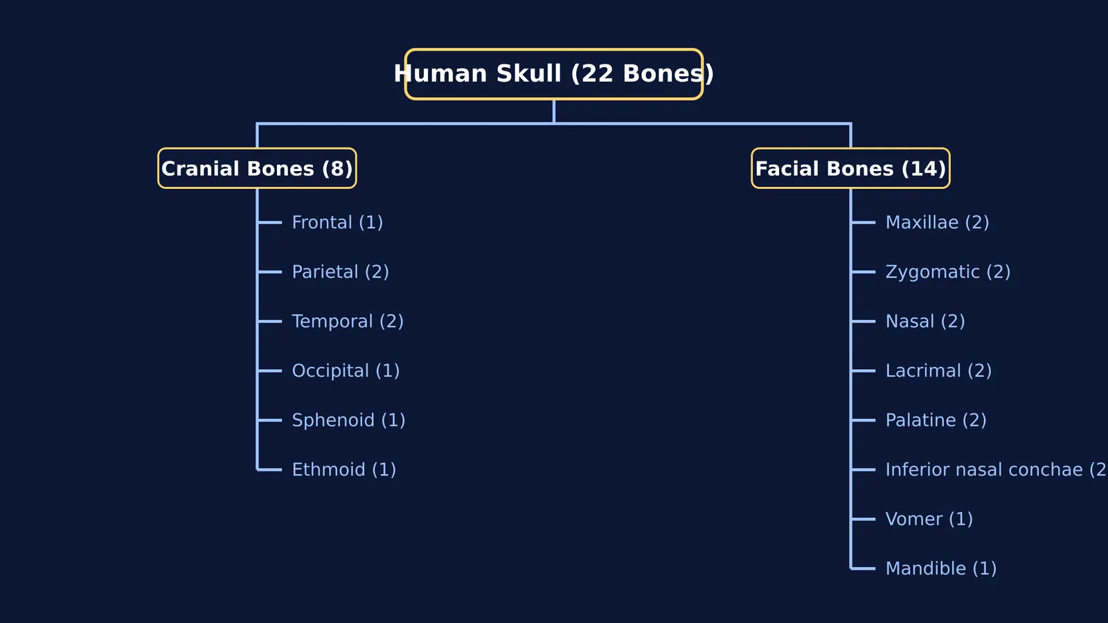
The 22 bones of the skull are split into 8 cranial bones and 14 facial bones 1.
The cranial bones form the neurocranium, a rigid, protective vault around the brain. The facial bones form the viscerocranium, the intricate framework that supports the facial muscles, the eyes, the nasal cavity, and the jaws.
What are the 8 cranial bones?
The neurocranium is built from eight bones that fuse together to encase the brain completely 1:
Frontal bone (1): Forms the forehead and the upper part of the eye sockets (orbits).
Parietal bones (2): Two large plates that form the roof and upper sides of the skull.
Temporal bones (2): Located on the lower sides of the skull, housing the delicate structures of the inner ear.
Occipital bone (1): Forms the back and base of the skull, containing a large opening (the *foramen magnum*) through which the spinal cord passes.
Sphenoid bone (1): A butterfly-shaped bone at the centre of the skull base that articulates with almost all other cranial bones.
Ethmoid bone (1): Sits between the eyes, forming part of the nasal cavity and the medial wall of the orbits.
What are the 14 facial bones?
The viscerocranium consists of 14 bones that determine the shape of the face and provide cavities for the sensory organs 1:
Maxillae (2): The paired bones that form the upper jaw and the front of the hard palate.
Zygomatic bones (2): The cheekbones, which also form the lateral walls of the eye sockets.
Nasal bones (2): Two small bones that meet in the middle to form the bridge of the nose.
Lacrimal bones (2): The smallest bones of the face, located on the inner walls of the eye sockets.
Palatine bones (2): L-shaped bones forming the back part of the hard palate and floor of the nasal cavity.
Inferior nasal conchae (2): Curved bony plates inside the nasal cavity that increase the surface area for warming and humidifying inhaled air.
Vomer (1): A thin, flat bone that forms the lower part of the nasal septum.
Mandible (1): The lower jawbone.
Which is the only movable bone of the skull?
Of the 22 bones in the skull, the mandible is unique: it is the only one that moves 1.
The mandible articulates with the temporal bone at the temporomandibular joint (TMJ). This complex joint, driven by powerful muscles of mastication (chewing) such as the masseter and temporalis, allows the jaw to elevate, depress, protrude, and move side-to-side. This mobility is essential for chewing, swallowing, and speech.
Every other bone in the skull is rigidly locked in place.
What are cranial sutures?
The immovable joints that lock the other 21 bones together are called sutures 1.
Sutures are made of strong fibrous tissue. In a newborn baby, these sutures are not fully fused. Instead, wide gaps of fibrous tissue called *fontanelles* (or "soft spots") separate the cranial bones. This flexibility serves two crucial purposes:
It allows the skull bones to overlap slightly during birth, helping the head pass through the birth canal.
It gives the brain room to expand rapidly during the first few years of life.
As the brain reaches its adult size, the fontanelles close, and the sutures gradually ossify (turn into solid bone), locking the skull into a single rigid structure. The most prominent sutures include the coronal suture (between the frontal and parietal bones), the sagittal suture (between the two parietal bones), and the lambdoid suture (between the parietal and occipital bones).
Learning anatomy with Fermi
The challenge of mapping the human body is ancient—dating back to Andreas Vesalius and the Fabrica: checking anatomy by hand—but the tools have evolved. Understanding the 3D relationships between these 22 bones can be challenging from text alone.
If you are studying human anatomy, the Fermi Concept Library covers every concept in biology. Instead of just answering questions, Fermi's AI tutor holds a line of reasoning across a session, deciding whether to explain the mechanics of the temporomandibular joint, ask you a question, or draw the anatomical figure on a board to check your understanding as it goes.


