On this page
An adult human body has roughly 206 bones, varying from the tiny stapes in your ear to the long femur in your leg. But if you were to cut any of these bones open, you would not find a single, uniform material. Instead, you would see two distinct types of osseous tissue working together: a dense outer shell and a porous inner core.
Understanding how these two tissues differ is a classic topic in histology and a frequent subject in NEET biology exams. Here is how their structures compare.
What is compact bone vs spongy bone structure?
The main difference between the two is their density and internal architecture.
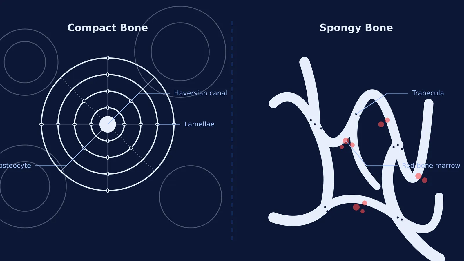
Compact bone (or cortical bone) is the hard, dense outer layer of every bone. It provides structural support and protection. In a healthy adult, the ratio of compact bone to spongy bone is approximately 80:20 by mass. Because it is so dense, compact bone must rely on a complex network of tiny canals to transport blood and nutrients to its living cells.
Spongy bone (also known as trabecular or cancellous bone) forms the inner layer. It is highly porous and looks like a rigid honeycomb. You will find spongy bone at the ends of long bones (the epiphyses), inside flat bones like the skull, and making up the bulk of the vertebrae, which lack a true cortex.
How does the Haversian system work in compact bone?
Compact bone is not a solid block of calcium. It is highly organised into functional cylindrical units called osteons, or Haversian systems.
Haversian canals: At the centre of every osteon is a Haversian canal, a vertical tunnel that carries blood vessels and nerves through the hard matrix.
Lamellae: Around this canal are concentric rings of mineralised bone matrix called lamellae, containing calcium hydroxyapatite crystals.
Lacunae and osteocytes: Between these rings are small spaces called lacunae. Mature bone cells, called osteocytes, reside inside these spaces and maintain the bone tissue.
Canaliculi: Tiny microscopic channels connect the lacunae to each other and to the central canal. This allows the osteocytes to share nutrients and communicate via gap junctions.
Because compact bone is so dense, this organised system is the only way to keep the bone cells alive. It is a defining feature of connective tissue types and examples you must know for biology exams.
Why is spongy bone lattice-like?
Spongy bone does not have osteons. Instead, it consists of a meshlike network of thin bony columns and plates called trabeculae.
This lattice structure serves two vital purposes:
Mechanical shock absorption: The trabeculae align along the lines of mechanical stress. When you jump or run, this network absorbs the impact from multiple directions, preventing the bone from fracturing.
Making bones lightweight: If your bones were entirely made of compact bone, your skeleton would be incredibly heavy, requiring massive muscles just to stand up.
The gaps between the trabeculae are not empty. They are filled with red bone marrow, the site of hematopoiesis (the formation of red and white blood cells).
How do bones change over time?
Bones are living, dynamic tissues that continuously change in response to mechanical stress and physiological demands. This lifelong process is known as bone remodeling.
Two key types of cells drive this process. Osteoblasts are the primary cells responsible for building new bone. They secrete a collagen-rich fluid called osteoid, which eventually mineralises to form the hard bone matrix. On the other hand, osteoclasts are large, multinucleated cells that break down and resorb old bone tissue.
How to study bone structure for exams
When preparing for exams like NEET, you will often need to differentiate between closely related anatomical terms, just as you would when studying the tendon vs ligament difference.
A good strategy is to sketch the cross-section of a long bone from memory. Draw the solid outer ring of compact bone and the porous inner network of spongy bone. If you are struggling to picture the microscopic differences, you can use the Fermi Concept Library to find the lesson on human histology and its place in the map. Being able to visualise where the osteoclasts and osteoblasts operate makes it much easier to understand how X-rays show bones inside the body.
By balancing the dense strength of compact bone with the lightweight flexibility of spongy bone through continuous remodeling, the human skeleton achieves an extraordinary feat of structural engineering.