On this page
Your inner ear turns vibrations into a wave in fluid, then turns that wave into electrical signals your brain can read. Every step depends on the one before, so one missing link sinks the answer. Here is the chain, followed by a frame for a 3-mark or 5-mark answer.
How does the cochlea convert sound into nerve signals?
The US National Institute on Deafness and Other Communication Disorders (NIDCD) describes the cochlea as "a snail-shaped structure filled with fluid" in its explainer How Do We Hear?. OpenStax Anatomy and Physiology 2e fills in the anatomy. Together:
Middle ear to oval window. The eardrum vibrates, and the malleus, incus and stapes amplify those vibrations and pass them to the cochlea through the oval window. More in our post on the ear ossicles and what each bone does.
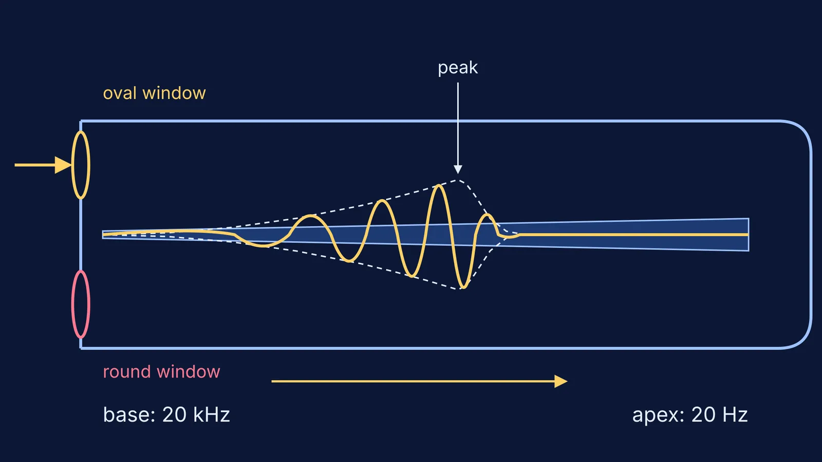
A wave in fluid. The oval window opens into the scala vestibuli, a fluid-filled tube running above the cochlear duct. The tube turns back towards the base as the scala tympani, which ends at the round window, an opening covered by a membrane. Vibrations at the oval window set up pressure waves in the fluid of both scalae.
The basilar membrane moves. The pressure waves move the basilar membrane, the floor of the cochlear duct. NIDCD calls this a travelling wave along the membrane.
Stereocilia bend. The organ of Corti sits on the basilar membrane. Its hair cells carry stereocilia, arranged from tallest to shortest, that reach up to the tectorial membrane. As the basilar membrane moves, the tectorial membrane slides across the stereocilia and bends them.
Ion channels open. When stereocilia bend towards the tallest one, tension in the protein tethers between them opens ion channels and the hair cell depolarises. Bend them the other way and the tethers slacken and the channels close.
The nerve fires. A stimulated hair cell releases an excitatory neurotransmitter at a synapse with a sensory neuron, as OpenStax Biology 2e puts it. The impulses travel along the cochlear branch of the vestibulocochlear nerve (the auditory nerve) to the brain.
Why do different parts of the cochlea hear different pitches?
OpenStax Biology 2e describes the basilar membrane as thicker, tauter and narrower near the base, and thinner, floppier and broader towards the apex. High frequencies make the membrane vibrate most near the entrance. Lower frequencies travel farther along before they peak. This is the place theory of pitch.
The human cochlea encodes frequencies between 20 and 20,000 Hz, with 20 Hz picked up at the apex and frequencies near 20 kHz at the base (OpenStax Anatomy and Physiology 2e). To find where your own hearing fades, try our human hearing range tone test.
What do inner and outer hair cells do?
In a 2008 review in Physiological Reviews, Jonathan Ashmore counts about 3,500 inner hair cells in each human cochlea (Ashmore, 2008; passage reproduced on BioNumbers). These cells connect to the auditory nerve and are considered the main sensory cells. The same review puts outer hair cells at about 11,000 per cochlea, in 3 or 4 rows. Outer hair cells act like motors: they produce forces driven by changes in their membrane potential, and the review traces the cochlea's amplification of sound to them.
In a board answer, one line each is enough: inner hair cells signal, outer hair cells amplify.
How do you write the cochlea answer in a board exam?
Use this five-part frame for any "explain the mechanism of hearing" question:
Entry: eardrum, then ossicles, then oval window.
Fluid: scala vestibuli, then scala tympani, ending at the round window.
Membranes: the basilar membrane moves, the tectorial membrane shears the stereocilia.
Transduction: ion channels open, the hair cell depolarises and releases neurotransmitter.
Signal: the auditory nerve carries impulses to the brain. Add place coding if the question mentions pitch.
Worked example. "Explain how the cochlea converts sound into nerve impulses. (5 marks)" Write one sentence per point in the frame, in order, and add a labelled cross-section showing the scala vestibuli, cochlear duct, scala tympani, organ of Corti, basilar membrane and tectorial membrane.
Before you submit, check for these common slips:
Writing that hair cells "hear" sound. They respond to bending.
Swapping base and apex. Base means high pitch, apex means low pitch.
Mixing up the coil with the membrane. The base is the wide end of the snail shape, where NIDCD places high pitches, but it is where the basilar membrane is narrowest.
Confusing the basilar membrane (under the hair cells) with the tectorial membrane (over the stereocilia).
If one term feels shaky, look it up in the Fermi Concept Library, where every biology concept has its own lesson and its place in the map. A travelling wave is also easier to see than to read, which is what the Fermi Simulation Lab is built for.
Next, see how your brain compares the two cochleas to work out which direction a sound comes from.