
On this page
Cartilage and bone are both connective tissues, and both keep their cells in small pockets called lacunae. After that, almost everything differs: the matrix, the cells, the blood supply, the healing speed and where each is found.
What is the main difference between cartilage and bone?
The short answer is minerals and blood vessels. Bone's matrix is mostly collagen fibres set in a mineralised ground substance containing hydroxyapatite, a form of calcium phosphate, according to the open textbook OpenStax Anatomy and Physiology 2e. Cartilage's matrix gets its distinctive character from something else: chondroitin sulfates that bind with proteins to form proteoglycans.
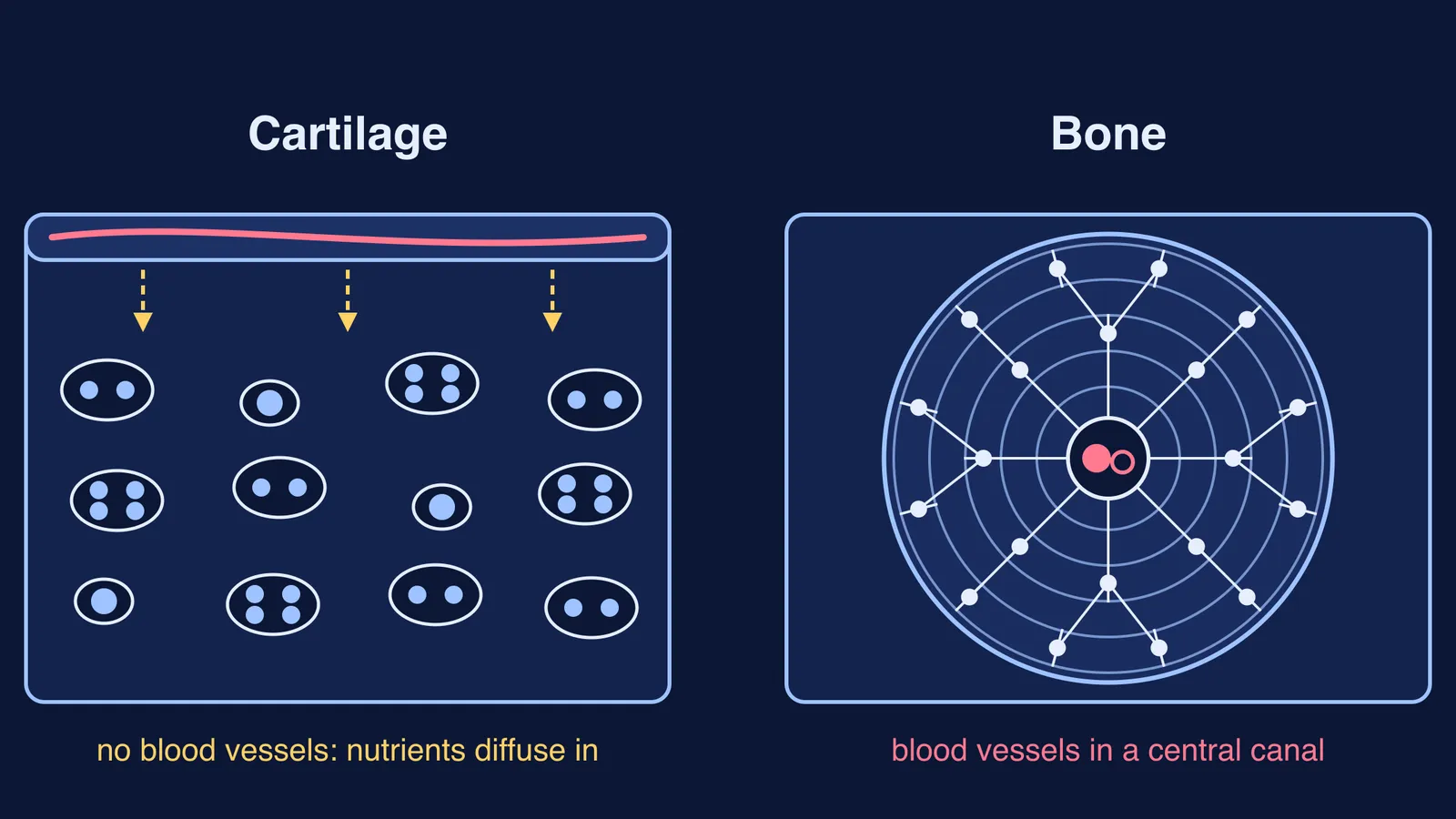
The same chapter states that cartilage is avascular, so nutrients diffuse through the matrix to its cells, which contributes to its very slow healing. Bone is highly vascularised and recovers from injury relatively quickly.
How do the matrix and cells of cartilage and bone differ?
Start with the matrix. By weight, bone is about 60% inorganic mineral, about 30% organic material and about 10% water, according to a review by Xu Feng in Current Chemical Biology (2009). The same review says about 90% of bone's organic part is type I collagen.
Articular cartilage, the hyaline cartilage that caps the ends of bones in joints, is up to 80% water by wet weight, according to a review by Sophia Fox, Bedi and Rodeo in Sports Health (2009). In that review, collagen makes up about 60% of the dry weight of cartilage, and proteoglycans account for 10% to 15% of the wet weight.
Those ingredients explain the difference in flexibility: hydroxyapatite crystals give bones hardness and strength, while collagen keeps them from being brittle, as OpenStax explains in its chapter on bone structure.
Now the cells. Cartilage cells are called chondrocytes, and they make up about 2% of the total volume of articular cartilage (Sophia Fox and colleagues). Bone has four cell types, all described in the OpenStax bone chapter:
Osteogenic cells: the only bone cells that divide.
Osteoblasts: form new bone.
Osteocytes: the most common cells of mature bone.
Osteoclasts: break bone down.
Why does cartilage have no blood supply, and why does it matter?
Articular cartilage is devoid of blood vessels, lymphatics and nerves, and has a limited capacity for intrinsic healing, according to the Sports Health review by Sophia Fox and colleagues. Cartilage is wrapped in the perichondrium, a layer of dense irregular connective tissue, as the OpenStax connective tissue chapter describes, and food for the chondrocytes has to travel through the matrix by diffusion.
Bone has its own plumbing. Arteries enter through openings in the shaft called nutrient foramina, central canals carry blood vessels and nerves, and tiny channels called canaliculi connect osteocytes with one another and eventually with the central canal, so nutrients arrive and wastes leave, according to the OpenStax bone structure chapter.
Where is cartilage found in the body, and where is bone?
The OpenStax connective tissue chapter and its chapter on cartilaginous joints give the main locations:
Hyaline cartilage, the most common type: rib cage, nose, the ends of bones at movable joints, the epiphyseal plate of a growing bone, and the costal cartilage that anchors the first rib to the manubrium of the sternum.
Fibrocartilage, made tough by thick bundles of collagen: the intervertebral discs between vertebrae and the pubic symphysis.
Elastic cartilage, which contains elastic fibres: the external ear.
Bone: the skeleton itself, with a dense outer layer and a lattice inside, which we compare in compact bone vs spongy bone.
The two tissues meet at joints. Articular cartilage there is 2 to 4 mm thick, according to the Sports Health review, and it reduces friction and acts as a shock absorber, according to OpenStax. For joint types, see our guide to ball and socket, hinge and pivot joints.
How can you remember cartilage vs bone for an exam?
On a blank page, answer these seven prompts for cartilage and for bone from memory, then check:
Matrix: proteoglycans and collagen, or mineral and collagen?
Cells: chondrocytes, or four bone cell types?
Where cells sit: lacunae in both.
Blood supply: none, or vessels in central canals?
Nutrients reach cells by: diffusion, or canaliculi?
Healing: very slow, or relatively fast?
Found in: three cartilage types with locations, or the skeleton?
Then try assertion-reason pairs. Assertion: cartilage heals more slowly than bone. Reason: cartilage has no blood vessels, so nutrients reach chondrocytes only by diffusion. Both are true, and the reason explains the assertion. Next: the intervertebral disc is hyaline cartilage. False; it is fibrocartilage.
For a wider drill, try our method to quiz the location and job of plant and animal tissues. If a term such as proteoglycan or lacuna keeps tripping you up, the Fermi Concept Library has every concept in biology with its own lesson and its place in the map.
Answer the seven prompts again in three days; reread the ones you missed.