
On this page
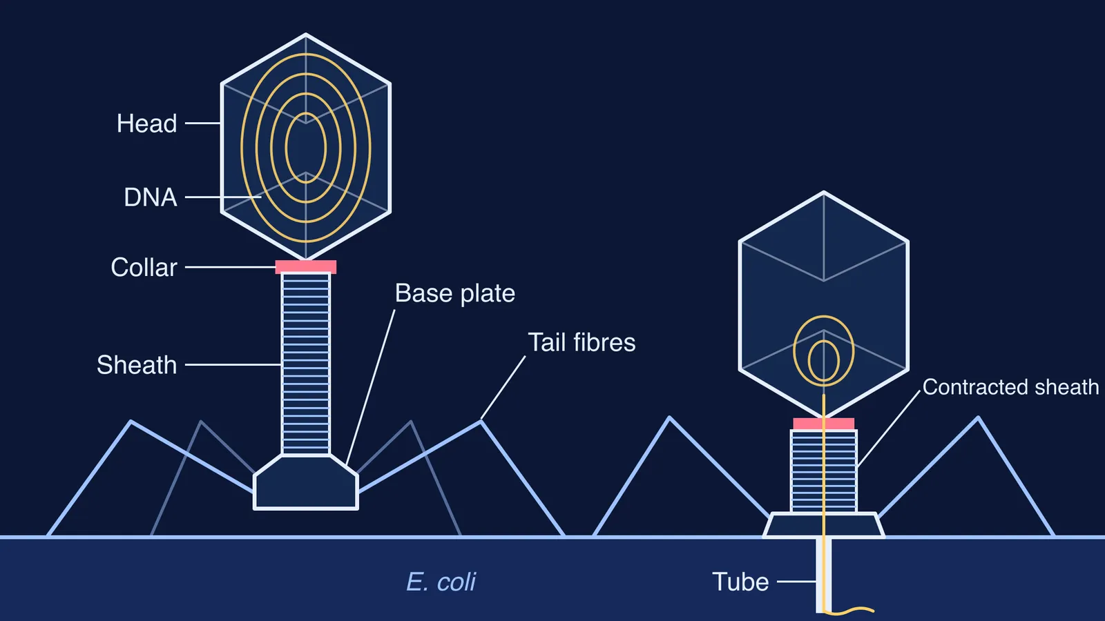
Your NCERT Class 11 Biology chapter on Biological Classification draws a bacteriophage in Figure 2.6b with four labels: head, collar, sheath and tail fibres (NCERT, Chapter 2). Exam questions often add a fifth part, the base plate, and ask what each part does. Here are the parts, their sizes in phage T4, and what each does during infection.
What are the parts of a bacteriophage T4?
Bacteriophage T4 infects *Escherichia coli*, and its measurements below come from a 2014 review by Moh Lan Yap and Michael Rossmann of Purdue University (Yap and Rossmann, Future Microbiology).
Head (capsid): a stretched icosahedron, 115 nm long and 85 nm wide, built mainly from 155 hexamers of one protein, gp23. It holds the 172 kbp double-stranded DNA genome.
Collar (neck): joins the head to the tail. The DNA enters and leaves the head through a portal protein at one corner (vertex) of the head. Whisker fibres on the neck help attach the long tail fibres during assembly and help the phage sense conditions suited to infection.
Sheath: a contractile tube made of 23 stacked rings of protein gp18, wrapped around a rigid inner tube. The whole tail is 92.5 nm long and 24 nm wide.
Base plate: a hexagonal, dome-shaped plate 52 nm across at the end of the tail. It anchors the fibres and carries a sharp tip with three lysozyme domains.
Tail fibres: six long fibres, each 145 nm, attached to the edge of the base plate and bent at a knee. Six short fibres stay folded underneath the base plate until the phage finds its host.
How does a bacteriophage infect a bacterium, step by step?
The same review describes a sequence in which each part acts in turn (Yap and Rossmann):
Find. The long tail fibres recognise the host. The tip protein, gp37, binds lipopolysaccharide on the outer surface of *E. coli*.
Signal. The base plate relaxes from a high-energy dome shape into a flatter star shape.
Grip. The short tail fibres unfold and bind the cell irreversibly.
Contract and drill. The sheath contracts, shortening to 42 nm long and 33 nm wide. The head turns by about 345 degrees, which pushes the sharp tip of the tail through the outer membrane like a drill, and the lysozyme domains break down the cell wall beneath it.
Inject. The DNA travels from the head, through the tube, into the cell.
The coat stays outside. In 1952, Alfred Hershey and Martha Chase showed with the related phage T2 that most of the phage DNA enters the cell on infection while most of the protein coat stays at the surface. They concluded that the coat is responsible for attachment and works as an instrument for injecting the DNA (Hershey and Chase, Journal of General Physiology). Phages can also carry bacterial genes between cells; see how Zinder and Lederberg discovered bacteriophage transduction.
Why does each part's shape match its job?
One line per label answers most "give the function" questions:
Head: a closed protein box, so it protects and stores the DNA.
Collar: a joint with a portal, so it connects head to tail and lets DNA pass.
Sheath: a spring of stacked rings, so it contracts and pushes.
Base plate: a hub with a sharp tip, so it switches shape on landing and starts the puncture.
Tail fibres: long, jointed legs with a binding tip, so they recognise the right host.
If you mix up viruses and bacteria in the same answer, our difference table for bacteria vs viruses sets the two side by side.
How do you draw a bacteriophage diagram for the exam?
Keep your sketch in proportion using the T4 measurements from Yap and Rossmann: the head is a little longer than the tail (115 nm against 92.5 nm), and each long fibre (145 nm) is longer than the tail itself. A quick drawing plan:
Draw a tall hexagon for the head and fill it with a coiled line for DNA.
Add a short, narrow collar under it.
Draw the sheath as a striped cylinder, slightly shorter than the head.
Put a flat hexagonal base plate at the bottom.
Draw the fibres from the base plate's edge, out and up, then bent down at a knee.
Label head, DNA, collar, sheath, base plate and tail fibres, with ruled lines that never cross.
Common mistakes: tail fibres drawn from the collar instead of the base plate, fibres drawn as straight spokes with no bend, and RNA written inside the head. To test yourself, use the cover-and-check method for textbook diagrams: hide the labels, rebuild them, then compare.
If viruses or capsids still feel shaky, the Fermi Concept Library gives every biology concept its own lesson and its place in the map. Fermi's AI tutor can draw the figure and check your understanding as it goes.