On this page
If you took PCB in Class 12 and want to work in a hospital without becoming a doctor, imaging technology is one of the clearest paths. It is a named profession in Indian law. The National Commission for Allied and Healthcare Professions Act, 2021 lists the Radiology and Imaging Technologist (diagnostic radiographer, MRI, CT, mammographer and diagnostic sonographer) under its Medical Radiology, Imaging and Therapeutic Technology category.
The same schedule describes the work: operating radiographic, ultrasound and other imaging equipment to produce images of body structures, under the supervision of a radiologist or other medical professional.
What does a BSc in radiology and imaging technology teach?
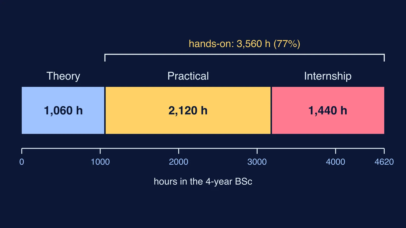
The Ministry of Health and Family Welfare's Model Curriculum Handbook for Medical Radiology and Imaging Technology sets out the BSc as 4 years or 8 semesters: 1,060 hours of theory, 2,120 hours of practical classes and 1,440 hours of internship, a total of 4,620 hours.
Do the arithmetic and the shape of the degree is clear. Practical classes are twice the theory hours, and with the internship, 3,560 of the 4,620 hours (about 77%) are hands-on. Expect to spend most of your time positioning patients, running equipment and checking images, not taking notes.
The handbook builds it up like this:
First semester: a foundation course covering India's healthcare system, communication, medical law and ethics, and patient safety.
Second semester: human anatomy and physiology, basic physics including radiological physics, and conventional radiography.
Third semester: clinical radiography and patient positioning, and modern imaging equipment.
By graduation: the final two semesters are a clinical internship, and the handbook expects graduates to carry out mammography, CT and MRI procedures independently and evaluate images for technical quality.
The handbook expects a Class 12 pass with Physics, Chemistry and Biology and an entrance test conducted by the university. Tests and cut-offs differ from one university to the next, so check the prospectus.
Which machines do radiology technologists run?
The Health Ministry handbook lists the imaging methods diagnostic radiographers use, including X-ray, fluoroscopy, CT, MRI, ultrasound, angiography, mammography and DEXA bone densitometry. Each works on different physics:
X-ray and fluoroscopy send X-rays through the body; fluoroscopy shows moving images in real time. Our explainer on how an X-ray shows bones covers why bone appears white.
CT aims a narrow X-ray beam that rotates quickly around the body, and a computer builds cross-sectional images from the signals, according to the US National Institute of Biomedical Imaging and Bioengineering.
MRI uses powerful magnets and radio-frequency pulses and no ionising radiation, but its field is strong enough to fling a wheelchair across the room, so metal objects are a serious safety concern around the scanner (NIBIB on MRI). See how an MRI scanner sees inside your body.
Ultrasound sends sound waves into the body and uses the echoes from tissue boundaries to build an image; it does not produce ionising radiation (NIBIB on ultrasound).
Every one of these machines runs on Class 11 and 12 physics: X-rays, magnetism or sound. If those feel abstract, the Fermi Simulation Lab has interactive simulations for ideas that are easier to see than to read, and the Fermi Concept Library gives each physics concept its own lesson.
Where do radiology and imaging technologists work?
The Health Ministry handbook lists hospitals, diagnostic and medical labs, clinical and medical research organisations, pharma and biotech companies, and medical equipment and device companies. Inside hospitals, diagnostic radiographers serve accident and emergency, outpatients, operating theatres and wards.
Job titles it names include radiographer, CT, MRI, mammography and cath lab technologist, applications specialist, PACS manager and radiological safety officer. It says BSc graduates are prepared to apply for the Level I Radiation Safety Officer role under Atomic Energy Regulatory Board (AERB) norms.
For teaching, the handbook notes that a Lecturer or Assistant Professor post needs a master's degree under UGC guidelines, and it recommends a master's programme of at least two years in the specialty.
How do you check a radiology course before you join?
Take these five questions to the admission office:
Where is the internship, and is it a full year in a working hospital?
Which machines will I operate myself, not just watch? The handbook recommends institutes have conventional and mobile X-ray, fluoroscopy, ultrasound with colour Doppler, multi-slice CT and mammography, with MRI preferably.
Is the college attached to a medical college or teaching hospital with enough patients?
How is radiation-safety training taught and assessed?
Where did last year's batch go: which hospitals, labs or companies?
Parents can try one line at the counselling desk: "Can we see the imaging department our child will train in?" A college that says no is telling you something.
Allied health professions now come under the NCAHP Act, so confirm the current regulations with the university before paying fees. For the wider picture, read our guide to allied health science courses after 12th PCB.