On this page
An MRI scanner does not photograph you; it listens. It puts your body in a strong magnet, sends in a short radio pulse, and records the faint radio signal that comes back from hydrogen nuclei in your tissues. If you know Class 12 magnetism and resonance, you already know much of the physics behind it.
Why does an MRI scanner look for hydrogen?
According to the Nobel Prize press release for the 2003 Physiology or Medicine prize, water makes up about two thirds of human body weight, and that high water content is why MRI works so well in medicine. Each water molecule has two hydrogen atoms, and the nucleus of each one is a single proton.
The release describes these nuclei as "microscopic compass needles". It also notes that different tissues hold different amounts of water, and that many diseases change water content, which then shows up in the image.
How does an MRI scanner see inside the body?
Line up the protons. A strong magnet "forces protons in the body to align with that field", as the US National Institute of Biomedical Imaging and Bioengineering (NIBIB) puts it. Medical scanners usually use 1.5 T or 3 T fields, while Earth's field is only 24 to 64 µT, so the scanner's magnet is tens of thousands of times stronger.
Tip them with a radio pulse. In a magnetic field the protons spin like wobbling tops (physicists call it precession) at a frequency set by the field strength. A radio pulse at exactly that frequency is absorbed. This matching is the resonance in "magnetic resonance", as the Nobel press release explains.
Listen as they relax. When the pulse stops, the protons fall back into line and give off radio waves. NIBIB notes that the time this takes, and the energy released, depend on the chemical surroundings, which is why different tissues give different signals.
Tell the signals apart by position. Extra "gradient" magnets make the field slightly stronger on one side, so each position gets its own frequency. Paul Lauterbur described imaging with gradients in Nature in 1973, and Peter Mansfield developed the idea further, showing how the signals could be analysed mathematically and how very fast imaging could be achieved; the two shared the 2003 Nobel Prize.
Build the picture. A computer sorts the mixed signal by frequency and turns it into slices, then a 3D image.
What radio frequency does an MRI scanner use?
The resonance rule is a straight line: frequency = γ × B, where γ is the proton's gyromagnetic ratio. The CODATA 2022 value published by NIST is 42.577 MHz per tesla. A worked example:
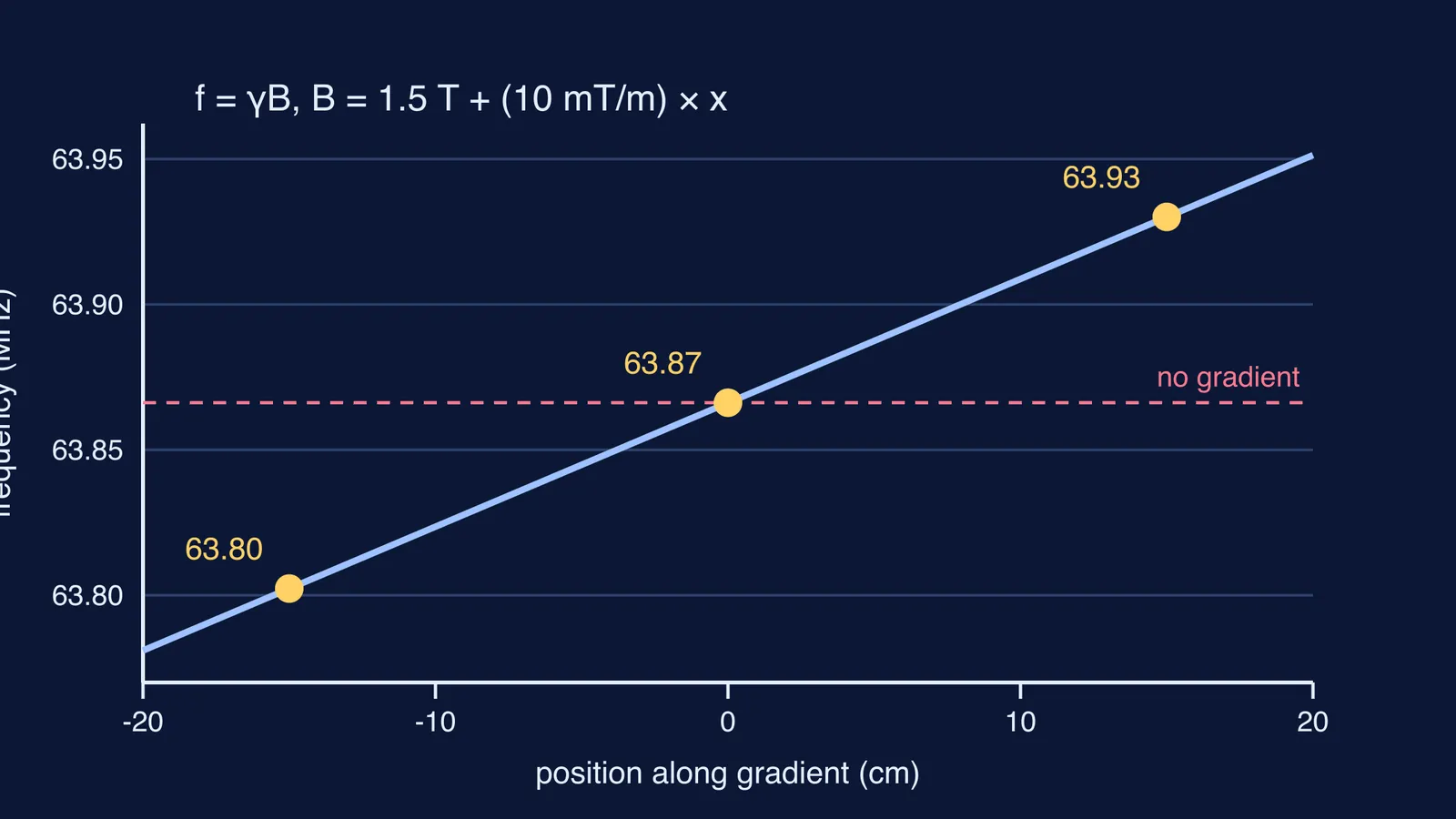
At 1.5 T: 42.577 × 1.5 = 63.87 MHz.
At 3 T: 42.577 × 3 = 127.73 MHz.
Now add a gradient. Suppose (for illustration) the field rises by 10 mT for every metre across the body, so 0.1 mT per centimetre. The frequency then changes by 42.577 × 0.0001 MHz = 4.26 kHz for every centimetre. Two points 15 cm apart differ by about 64 kHz. That small difference tells the computer where each signal came from; the diagram above plots this line. If you have met series LCR resonance, the tuning idea is the same.
Is an MRI scan safe, and why is it so loud?
The US FDA says MR images are made "without using any ionizing radiation". That is the big difference from how X-rays show bones. The FDA also lists real risks: the magnet attracts metal objects such as keys and phones, which can become projectiles, the radio energy could warm the body, and the changing magnetic fields make loud knocking noises.
NIBIB notes the sound can reach up to 120 decibels in some scanners, which is why ear protection may be needed. The FDA's MRI page says a typical scan lasts 20 to 90 minutes, depending on the part of the body being imaged. The magnet is also why staff ask about metal and implants before a scan.
How do you explain MRI in an exam or to your family?
Use this five-link chain, one line per link.
Body is mostly water; water has hydrogen; each hydrogen nucleus is a proton that acts like a tiny magnet.
Strong field B lines the protons up and sets them precessing at f = γB.
A radio pulse at f is absorbed (resonance) and tips them.
As they relax they emit radio waves; different tissues relax differently.
Gradients make f depend on position, so the signal can be mapped into an image.
Precession is the hardest step to picture; a wobbling spinning top is the same motion. The Fermi Simulation Lab has interactive simulations for the ideas that are easier to see than to read. For the history, read how Edward Purcell and Felix Bloch discovered NMR.