On this page
In 1946, two teams of physicists on opposite sides of the United States made a discovery that would eventually sit in every major hospital in the world. Edward Purcell at Harvard and Felix Bloch at Stanford independently proved that nuclear magnetic resonance (NMR) could be detected in ordinary chunks of matter, not just in a vacuum. Their breakthrough opened a new window into the atomic structure of solids and liquids.
What is nuclear magnetic resonance?
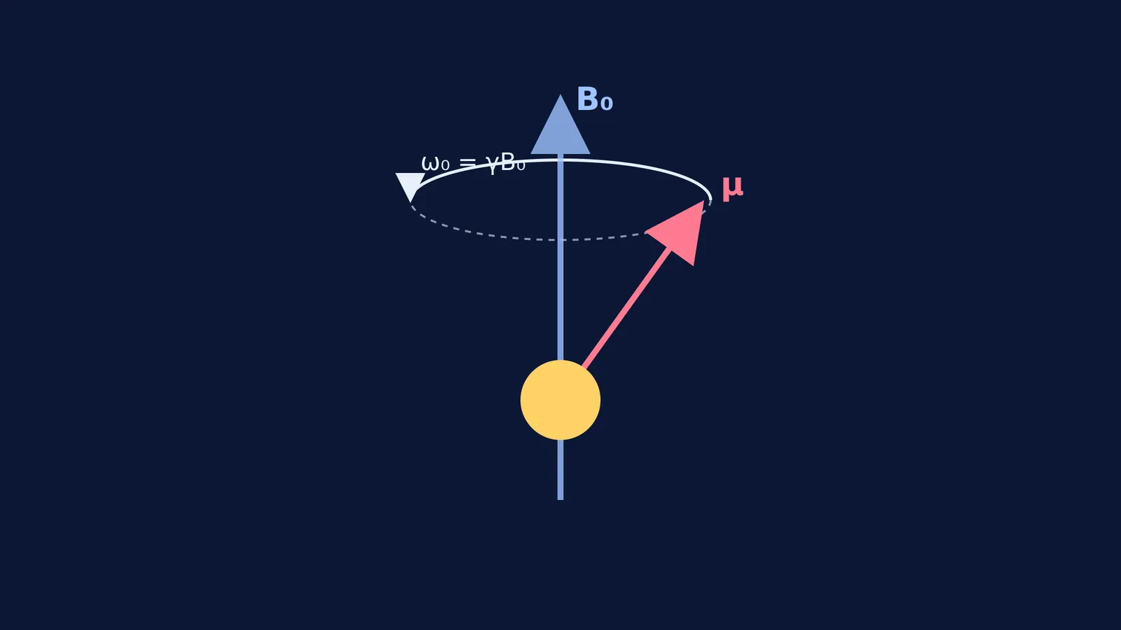
Atomic nuclei with an odd number of protons or neutrons have a property called spin. Because protons have a positive electrical charge, this spinning motion makes them behave like tiny bar magnets with a north and south pole. When placed in a strong external magnetic field (which you can explore in our guide on how to visualise the magnetic field of a solenoid), these nuclear magnets align either parallel or anti-parallel to the field.
If you hit them with a radio-frequency pulse at exactly the right frequency—the resonant frequency—they absorb the energy and flip their alignment to the higher energy state. When the pulse ends, they relax back to their original state, releasing their own faint radio signal in the process. This cycle of absorption and emission is called nuclear magnetic resonance.
How did Edward Purcell detect NMR in a solid?
Before 1946, physicist Isidor Rabi had successfully demonstrated NMR by sending beams of vaporised molecules through a vacuum. Physicists did not yet know if the resonance effect could be seen in solid or liquid matter. In everyday bulk matter, atoms are packed tightly together and constantly colliding, which could disrupt the delicate magnetic alignment.
In late 1945, Edward Purcell, working with Henry Torrey and Robert Pound at Harvard University, set out to test a solid block of paraffin wax. Paraffin is a hydrocarbon, meaning it is extremely rich in hydrogen atoms (protons). They placed the wax sample in a strong electromagnet and slowly swept a radio-frequency signal through it.
They found that the wax absorbed a measurable amount of the radio energy at one specific frequency, proving that the tightly packed protons were resonating together. Their paper, Resonance Absorption by Nuclear Magnetic Moments in a Solid, was published on 1 January 1946.
How did Felix Bloch detect NMR in water?
At the exact same time, Felix Bloch, working with William Hansen and Martin Packard at Stanford University, ran a similar experiment on a liquid. They placed a small spherical bulb of water inside a strong magnetic field.
Instead of measuring the direct absorption of energy like Purcell's team, Bloch's group wrapped a receiver coil around the water sample. When the protons resonated and precessed (wobbled like a spinning top), their moving magnetic fields induced a faint oscillating voltage in the receiver coil. This relied on the same basic principle as Faraday's discovery of electromagnetic induction in 1831.
Bloch called this method "Nuclear Induction", and the paper was published on 1 February 1946. For proving that NMR works in everyday matter, Purcell and Bloch shared the 1952 Nobel Prize in Physics.
When you study electromagnetism, picturing invisible fields and how they affect matter is difficult. The Fermi Simulation Lab lets you change the strength of a magnetic field and see the effects in real time, making these abstract physics ideas easier to see than read.
Why does NMR matter for MRI scanners?
Purcell and Bloch's discovery of NMR in bulk matter is the direct physical basis of Magnetic Resonance Imaging (MRI). An MRI scanner is essentially a large, advanced NMR machine designed for human bodies.
The human body is mostly composed of water and fat, both of which contain vast amounts of hydrogen protons. When a patient goes inside an MRI scanner, the machine's large superconducting magnet aligns their protons. The scanner then sends targeted radio pulses to flip them.
As the protons relax, they emit a radio signal. Because protons in different environments (such as a dense bone, a soft muscle, or a fluid-filled tumour) take different amounts of time to relax back to normal, the scanner can use these distinct signals to map the inside of the body in high contrast. This provides detailed medical images without using harmful ionising radiation like X-rays.
How to check a physics claim?
If you are learning about a major physics discovery, you can often trace it back to the original paper to separate facts from myths:
Find the authors: Note the names of the key physicists involved (e.g., Edward Purcell, Felix Bloch).
Find the year: Look for the year of the initial breakthrough (1946).
Search the journal: Many foundational physics papers of the 20th century were published in *Physical Review*.
Read the abstract: The abstract will clearly state the material tested (such as paraffin wax or water) and the experimental method used.
Tracing a discovery back to its primary source allows you to learn the exact facts and understand how the experiment was actually done.