On this page
Every school microscope practical has the same moment. You find the cells at low power, click to the 40× objective, and suddenly the view is dark, most of the cells are gone, and the one you wanted has slid out of sight. None of that is bad luck. All three changes follow from two short formulas, and you can predict them before you turn the nosepiece.
How does magnification change the field of view?
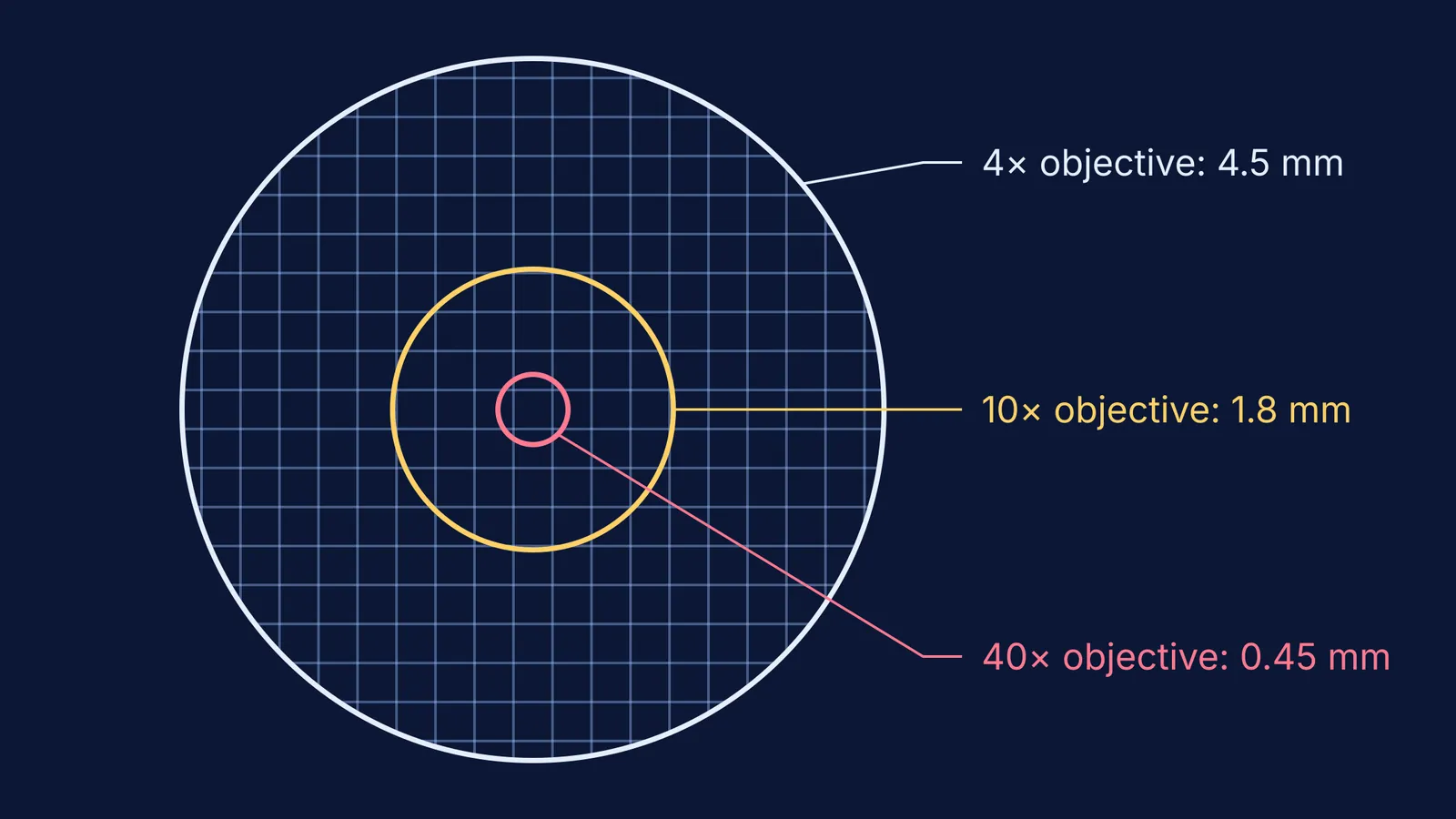
The field number is the diameter of the view field in millimetres, measured at the intermediate image plane inside the microscope (Nikon MicroscopyU). The field of view on your slide is the field number divided by the objective magnification (Nikon MicroscopyU). A 10× eyepiece typically has a field number between 16 and 18 mm, according to the same page.
Take an 18 mm field number and work through the three objectives found on many school microscopes:
4× objective: 18 ÷ 4 = 4.5 mm across
10× objective: 18 ÷ 10 = 1.8 mm across
40× objective: 18 ÷ 40 = 0.45 mm, which is 450 µm across
The eyepiece's magnification makes the image look bigger, but only its field number enters this formula. The objective sets how much of the slide fits in the circle.
Why do you see fewer cells at higher magnification?
The number of cells you can count depends on the area of the circle, not its width. The width is field number ÷ objective magnification (Nikon MicroscopyU), and area goes with the diameter squared, so going from 10× to 40× cuts the diameter by 4 and the area by 4 × 4 = 16.
That gives you a prediction you can check. If you count about 160 cells in the 10× field of an onion peel, expect roughly 10 at 40×. If your 40× count comes out much higher than that, you may be counting cells only partly in view, so set a rule first (count cells touching the top and left edge, skip the bottom and right).
Why does the image get darker at high power?
In transmitted light, image brightness is proportional to (NA/M)², where NA is the objective's numerical aperture and M is its magnification (Evident Scientific). Higher-power objectives usually have a larger NA, which only partly makes up for the loss.
Evident's own table shows this. A 10× plan achromat with NA 0.25 scores 6.25 on their transmitted-brightness scale, while a 40× plan achromat with NA 0.65 scores 2.64, well under half as bright (Evident Scientific). So when you switch up, open the diaphragm or raise the light before you decide the slide is the problem.
How do you estimate cell size from the field of view?
This needs no stage micrometer, only the field number printed on your eyepiece.
Work out the field diameter for your objective: field number ÷ objective magnification (Nikon MicroscopyU).
Convert it to micrometres: 1 mm = 1000 µm.
Count how many cells fit end to end across the diameter of the circle.
Divide: cell size ≈ field diameter ÷ number of cells across.
Check at a second objective. A real cell should come out about the same size at both.
Worked example: at 10× with an 18 mm eyepiece, the field is 1.8 mm, or 1800 µm. If 8 onion cells fit along the diameter, each is about 1800 ÷ 8 = 225 µm long. Switch to 40×: the field is 450 µm, so you should now see about 2 cells across. If you see 2, your estimate holds.
You can also run it backwards as a sanity check. Human red blood cells measure about 7.3 µm across (BioNumbers), so roughly 60 of them would line up across a 450 µm field at 40×. How much detail you can resolve is a separate limit, covered in why resolution matters in light and electron microscopes.
A five-question prediction sheet
Before your next practical, write down your answers, then test each one:
What is the field diameter at 4×, 10× and 40× for your eyepiece?
By what factor will the cell count drop from 10× to 40×?
Will the image be brighter or dimmer at 40×, and what will you adjust?
How many cells will fit across the field at 40×, given your 10× count?
What cell size do you get at each objective, and do they agree?
The Fermi Simulation Lab has interactive simulations for the ideas that are easier to see than to read. If the optics itself is the shaky part, our guide to what to watch in a convex lens image formation simulation covers how a lens forms a magnified image, and the limits of physics simulations explains why the real microscope on the lab bench still matters.
Next practical, count the cells at 10×, write down how many you expect at 40×, and only then turn the nosepiece.