On this page
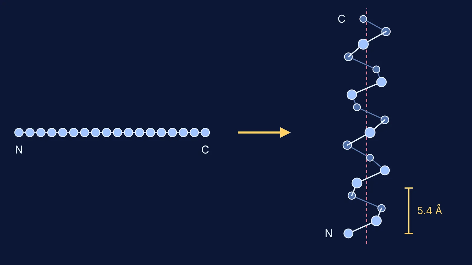
A protein starts as a string of amino acids, and its job depends on the 3D shape that string folds into. For decades, finding that shape meant painstaking lab work. Then an AI model called AlphaFold2 learned to predict it from the amino acid sequence, and its makers shared a Nobel Prize.
Why is predicting a protein's shape so hard?
The 2024 Nobel Prize in Chemistry announcement explains that amino acids link into long strings that fold into a three-dimensional structure, and that structure is "decisive for the protein's function". Researchers had been trying to predict structures from sequences since the 1970s.
According to the AlphaFold2 paper in Nature (Jumper and colleagues, 2021), experiments had solved the structures of around 100,000 unique proteins, a small fraction of the billions of known sequences. Solving a single structure can take months to years.
How did AI solve protein folding?
Strictly, AI solved one part: predicting the final shape from the sequence, which the Nature paper calls "the structure prediction component of the 'protein folding problem'". AlphaFold2, a deep-learning neural network, combines physical and biological knowledge about protein structure with multiple sequence alignments: your protein's sequence lined up against related versions from other organisms, showing which positions stay the same.
The test was CASP14, a round of the blind-prediction experiments run by the Protein Structure Prediction Center. AlphaFold2's predictions were competitive with experimental structures in a majority of cases, and well ahead of other methods (Jumper et al., 2021).
The Nobel committee says AlphaFold2 has been used to predict the structure of virtually all of the 200 million proteins researchers have identified, and more than two million people from 190 countries have used it. Demis Hassabis and John Jumper shared half the prize "for protein structure prediction". The AlphaFold Protein Structure Database makes more than 200 million of these predictions free to anyone.
Why does it matter for medicines?
Many medicines work by binding to a protein, so its shape gives chemists a place to start. The Nobel committee notes that predicted structures are helping researchers understand antibiotic resistance and picture enzymes that break down plastic.
AlphaFold 3 (Abramson and colleagues, Nature, 2024) goes further and predicts complexes: proteins together with nucleic acids (DNA and RNA), small molecules and ions. For proteins bound to small molecules, it was far more accurate than state-of-the-art docking tools. A predicted shape is a starting point for drug design, not a drug.
What can't AlphaFold tell biologists?
Every prediction comes with a confidence score called pLDDT, from 0 to 100. EMBL-EBI's AlphaFold course explains that above 90, the backbone and side chains are usually accurate; above 70, the backbone is usually right but some side chains may be misplaced. Below 50, the region may be naturally floppy (disordered), or the model may lack the information to predict it.
A study in Nature Methods by Terwilliger and colleagues compared predictions with experimental maps. Many matched remarkably closely, but even some very high-confidence predictions were distorted or had domains oriented the wrong way. Predictions also ignore bound molecules, chemical modifications and surroundings. The authors suggest treating them as "exceptionally useful hypotheses" and checking structural details by experiment.
A 2023 PLOS ONE study by Pak and colleagues found very weak or no correlation between AlphaFold's confidence metrics and how single mutations changed a protein's stability.
Worked example: what does AlphaFold say about haemoglobin?
Try this with human beta-globin, the haemoglobin chain behind sickle cell disease.
Look up entry AF-P68871-F1 in the AlphaFold database. The chain is 147 amino acids long, its mean pLDDT is 97.19, and 97.3% of residues are in the very high band. One normal chain is predicted very confidently.
Add the biology. MedlinePlus Genetics explains that adult haemoglobin usually has four subunits, two beta and two alpha, each bound to an iron-containing heme. In sickle cell disease, glutamic acid is replaced by valine at position 6 (Glu6Val).
Ask what the prediction misses. In sickle cell, haemoglobin S subunits "stick together" into long, rigid molecules (MedlinePlus). A prediction of one chain shows neither the heme nor that sticking, and AlphaFold's scores are a weak guide to a single mutation's effect (Pak et al., 2023).
Ask these four questions of any predicted structure:
How confident is each region (pLDDT)?
Does the protein work alone, or with partners and bound molecules?
Is my question about one sequence, or about a mutation?
Has an experiment confirmed the detail I care about?
How should a biology student study this?
Our guide to protein structure from primary to quaternary walks through each level, and point and frameshift mutations explains why one changed letter can matter. For the 3D side, see chemistry tools for seeing molecules in 3D.
If the jump from sequence to shape still feels abstract, the Fermi Concept Library has a lesson for every concept in biology and chemistry. Fermi's AI tutor works out what you already know and builds a plan for the session, so you know where to begin.