
On this page
In the early 1650s Thomas Bartholin (1616 to 1680), a Danish anatomist traced thin vessels in human and animal bodies. What he found helped take away a job the liver had held in medicine for centuries. If you are studying lymph in Class 11 biology, his work sits behind one line in your textbook: the lymphatic system collects tissue fluid and drains it back to the major veins.
Who discovered the lymphatic system?
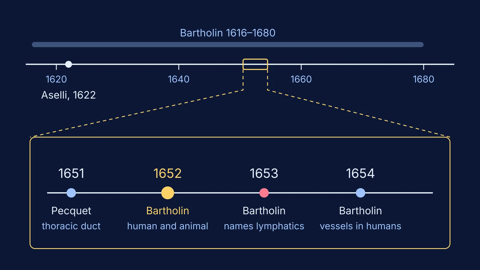
No single person did. It was pieced together across the 17th century. A 2017 history in the Journal of Anatomy by Natale, Bocci and Ribatti gives the order:
1622: Gaspare Aselli, dissecting a well-fed dog, saw "several thin and beautiful white cords". These were the lacteals, named for the milk-like fluid they carried.
1651: Jean Pecquet described the thoracic duct and the reservoir of the chyle, and showed that the lacteals empty there rather than into the liver.
1652: Bartholin published De lacteis thoracicis in homine brutisque, his observations in humans and animals. This first book still followed Galen's doctrine.
1653: His second book, Vasa lymphatica, named the clear-fluid vessels "lymphatics" for the first time.
A 2017 review in the European Journal of Anatomy by Sanjib Kumar Ghosh fills in the human part. Bartholin dissected human bodies and found vessels carrying clear fluid, unlike the cloudy chyle in the lacteals. These vessels did not enter the liver; they carried their contents away from it and into the blood. In 1654 he published Vasa Lymphatica in Homine Nuper Inventa, on lymphatic vessels newly found in humans.
So the short answer: Aselli found the lacteals, Pecquet traced them to the thoracic duct, and Bartholin is credited with describing and naming the lymphatic vessels in humans. Natale and colleagues note that who came first is still debated.
Why did Bartholin hold a funeral for the liver?
For about 15 centuries, medicine accepted Galen's teaching, as the Journal of Anatomy history notes. In Galen's view, the Ghosh review explains, venous blood was made in the liver.
Bartholin's 1653 title ended with "hepatis exsequiae", the funeral rites of the liver. Natale and colleagues read it as a witty epitaph for the idea that chyle moved to the liver. He had shown that the milky lacteals and the new clear vessels both reached the thoracic duct, not the liver, and he denied that the liver was where blood was made.
Ghosh adds that by 1654 Bartholin had come to see the lacteals and the vasa lymphatica as parts of one system, with fluids that differed in fat content.
Was it Bartholin or Rudbeck?
Olof Rudbeck, a young Swedish medical student, made similar observations at almost the same time. Ghosh writes that Rudbeck is generally accepted as the first to demonstrate the lymphatics in humans, at the royal court of Sweden in 1652, while Bartholin is credited as the first to publish.
Rudbeck later wrote a 752-word Latin letter charging Bartholin with pre-dating a key observation. C. T. Ambrose translated it in the Scandinavian Journal of Immunology in 2007 and argues that, by raising the question of priority, the letter stirred interest in the lymphatic system and hastened its study.
What does Class 11 say about lymph?
The NCERT Class 11 Biology chapter Body Fluids and Circulation covers it in section 15.2, Lymph (Tissue Fluid). Each point links back to the history:
As blood passes through capillaries, water and small water-soluble substances move into the spaces between cells. The larger proteins and most formed elements stay behind. This is tissue fluid, with the same mineral distribution as plasma.
The lymphatic system collects this fluid and drains it to the major veins. That is Bartholin's route, which Ghosh traces through the thoracic duct into the left subclavian vein.
Lymph is colourless: Bartholin's "clear fluid", as opposed to cloudy chyle.
Lymph carries lymphocytes for immune responses, plus nutrients and hormones.
Fats are absorbed through lymph in the lacteals of the intestinal villi: Aselli's white cords.
How can you revise lymph using this story?
Try this 10-minute plan the next time you revise Body Fluids:
Minutes 0 to 2: draw the route from memory. Blood capillary, tissue fluid, lymph vessel, thoracic duct, major vein. Mark where the lacteals join.
Minutes 2 to 5: answer aloud. Why is lymph colourless while chyle looks milky? What stays inside the capillary when tissue fluid forms?
Minutes 5 to 8: fix one claim. Suppose a classmate says tissue fluid is just plasma that leaked out. What is missing? The larger proteins and most formed elements stay in the vessels, though the minerals match plasma.
Minutes 8 to 10: tell the story in three sentences with three names: Aselli, Pecquet, Bartholin.
If plasma, tissue fluid and lymph still blur together, the Fermi Concept Library has a lesson for every concept in biology, each with its place in the map. If you are not sure where your gaps are, Fermi's AI tutor works out what you already know and builds a plan for the session.
Start with the drawing in step 1.