On this page
When Francis Crick proposed the central dogma, he faced a puzzle: amino acids have no chemical affinity for nucleic acid templates. In 1958, Crick formulated the adaptor hypothesis, predicting that dedicated RNA adaptors must carry amino acids to messenger RNA codons. In 1965, biochemist Robert Holley isolated and sequenced the first transfer RNA (tRNA) from yeast, proving Crick's hypothesis and revealing its classic cloverleaf secondary structure.
Why did biology need an adaptor molecule?
In the late 1950s, the connection between genes and proteins was an unsolved question. While DNA stored sequence information in four bases, protein synthesis required assembling twenty distinct amino acids into precise polypeptide chains, as Francis Crick noted when formulating the adaptor hypothesis.
Amino acid side chains differ widely in charge, polarity, and size, but none possess hydrogen-bonding surfaces that specifically recognise adenine, cytosine, guanine, or uracil. Crick argued that amino acids could not bind mRNA templates directly; instead, each amino acid attaches to a dedicated adaptor molecule made of RNA that carries the amino acid at one site and matches codons on mRNA through complementary base-pairing at another.
How did Robert Holley sequence alanine tRNA?
Determining an RNA sequence in the 1960s required new separation methods. Robert Holley spent nine years at Cornell University developing techniques to isolate and sequence yeast alanine tRNA, as recounted in his 1968 Nobel lecture.
Because individual tRNAs make up a tiny fraction of total cellular RNA, Holley used countercurrent distribution to isolate 1 gram of purified alanine tRNA from 140 kilograms of commercial baker's yeast, as detailed in the same Nobel lecture.
His team cleaved the purified RNA with two enzymes:
Pancreatic ribonuclease, which cuts after pyrimidine bases (U and C).
T1 ribonuclease, which cuts after guanosine (G).
By carrying out partial digestion with T1 ribonuclease at 0 degrees Celsius, Holley cleaved the molecule at a single exposed site into two halves. Overlapping these fragments allowed Holley to establish the complete 77-nucleotide sequence in Science on March 19, 1965. Holley shared the 1968 Nobel Prize in Physiology or Medicine with Har Gobind Khorana and Marshall W. Nirenberg, whose work with synthetic RNA polymers made by Har Gobind Khorana and Marshall Nirenberg's cell-free translation assays established the genetic code.
What does the tRNA cloverleaf structure look like?
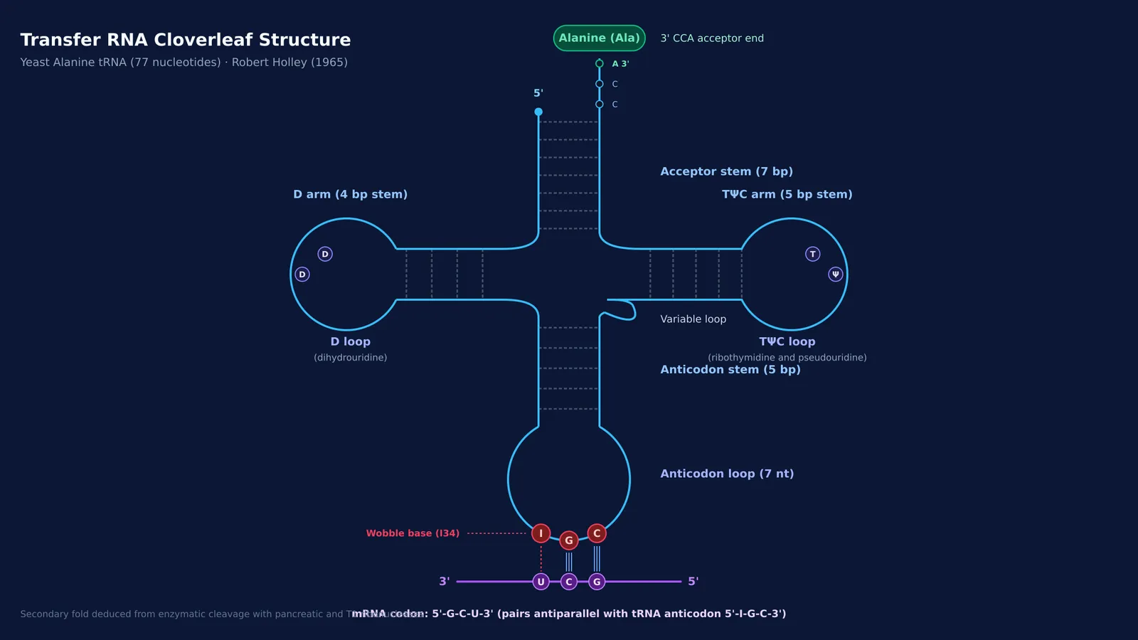
Analyzing the 77-nucleotide sequence in Science revealed internal complementary segments that pair into four stems and three loops, forming a cloverleaf:
Acceptor stem: A 7-base-pair stem ending in the unpaired sequence 5'-CCA-3', where the amino acid attaches to the terminal adenosine.
D-arm: A stem and loop containing the modified base dihydrouridine.
Anticodon arm: A 5-base-pair stem with a 7-nucleotide loop displaying the anticodon triplet (5'-IGC-3' in yeast alanine tRNA).
TΨC arm: A stem and loop containing ribothymidine, pseudouridine, and cytidine.
Variable loop: A short loop connecting the anticodon and TΨC arms.
While the cloverleaf shows the two-dimensional base pairs, X-ray crystallography by Alexander Rich and colleagues in 1974 proved that tRNA folds in three dimensions into an inverted L-shaped structure. This tertiary fold places the 3' CCA amino acid attachment site approximately 80 angstroms away from the anticodon loop.
How does tRNA read codons during protein synthesis?
During protein synthesis, tRNA serves as an adaptor by linking codons to amino acids, ensuring accurate reading codons without mistakes during translation.
This fidelity relies on two checkpoints:
Aminoacylation: An aminoacyl-tRNA synthetase links the correct amino acid to the 3' CCA tail.
Antiparallel pairing: Inside the ribosome, the tRNA anticodon pairs antiparallel with the mRNA codon.
Because alanine tRNA has inosine at position 1 of its anticodon (5'-IGC-3'), it can pair with U, C, or A at the third codon position through wobble pairing, as formulated in Francis Crick's wobble hypothesis80022-0). This allows a single tRNA to read three separate alanine codons: 5'-GCU-3', 5'-GCC-3', and 5'-GCA-3'.
How to decode codon-anticodon pairing step by step?
To determine which codons a tRNA reads, follow this four-step procedure:
Identify the anticodon (5' to 3'): For yeast alanine tRNA, the anticodon sequence is 5'-I-G-C-3' (nucleotides 34 to 36).
Align the mRNA codon antiparallel (3' to 5'): Because pairing in the ribosome is antiparallel, the 5' anticodon base aligns with the 3' codon base, and the 3' anticodon base aligns with the 5' codon base.
Determine Watson-Crick pairs at positions 2 and 3: The 3' anticodon cytosine (C36) pairs strictly with 5' codon guanine (G1), and the middle guanine (G35) pairs with middle cytosine (C2), establishing the codon prefix 5'-G-C-…-3'.
Apply wobble rules at position 1 (5' I): Inosine (I34) at the wobble position pairs with uracil (U), cytosine (C), or adenine (A) at the 3' end of the codon, recognizing three distinct codons: 5'-G-C-U-3', 5'-G-C-C-3', and 5'-G-C-A-3'.
Visualizing how single RNA strands fold into stems and loops can be difficult on paper. The Fermi Simulation Lab provides interactive simulations for the ideas that are easier to see than to read. To explore molecular genetics in depth, the Fermi Concept Library covers every concept in physics, chemistry, biology, mathematics and computing, each with its own lesson and its place in the map, while the Fermi Problem Library offers worked problems with a step-by-step solver that explains the reasoning rather than printing an answer.