On this page
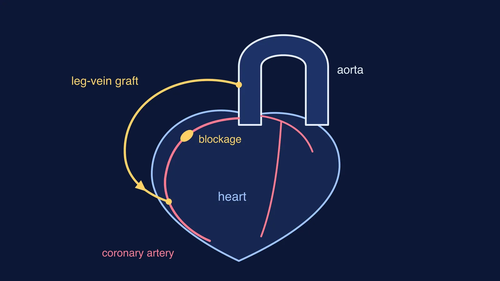
The heart's own muscle gets its blood through the coronary arteries, which branch off the aorta at its base. In 1967 René Favaloro, an Argentine surgeon working in the United States, showed that a piece of vein from a patient's own leg could carry blood past a blocked stretch of one of those arteries.
What did René Favaloro do in 1967?
Favaloro was born in La Plata, Argentina, in 1923, spent 12 years as a country doctor in La Pampa, and went to the Cleveland Clinic in 1962, according to a tribute by surgeon Denton Cooley in the Texas Heart Institute Journal. There he studied the thousands of coronary X-ray films (cineangiograms) made by Mason Sones.
In May 1967 he operated on a 51-year-old woman whose right coronary artery was totally blocked in its first third (Cooley, 2000). A historical review in the Malta Medical Journal describes that first operation as a saphenous vein graft interposition: a length of leg vein set into the artery in place of the diseased segment. About a week later Sones imaged the patient and showed the graft was open (Hektoen International; Cooley gives eight days).
The same Malta review says these operations paved the way for the aorto-coronary saphenous vein bypass in October 1967: one end of the vein sewn to the aorta, the other to the coronary artery beyond the blockage. Favaloro did 13 more cases by October and published his results in April 1968 (Rocha, 2017) in The Annals of Thoracic Surgery66351-5).
How does a bypass get blood past a blocked artery?
Coronary heart disease is when the heart's arteries cannot deliver enough oxygen-rich blood to the heart muscle because plaque has narrowed them, as the US National Heart, Lung, and Blood Institute defines it.
Coronary artery bypass grafting connects healthy blood vessels from elsewhere in the body to vessels above and below the blocked artery, and these are usually arteries from the arm or chest or veins from the legs, according to NHLBI's page on CABG.
Favaloro's idea came from work his team already knew. Cleveland surgeons had used saphenous vein grafts to rebuild kidney and leg arteries, and he later recalled asking, "Why not use it at the coronary level?" (Captur, 2005). For a refresher on how blood reaches the heart muscle itself, read our explainer on the hepatic portal system and coronary circulation.
Was Favaloro the first to do coronary bypass surgery?
It depends on what you count. A 2017 review by Eduardo Rocha lists earlier work: Robert Goetz operated with a tantalum ring in 1960 and published the first human coronary bypass in 1961, Vasilii Kolesov published his Russian experience in 1965, and Edward Garrett's team did a saphenous vein graft in November 1964 that was not reported until 1973.
Rocha concludes that who invented the procedure is debatable, but Favaloro marks the start of modern bypass surgery. By December 1968 his team they had done a double bypass to the right coronary artery and the left anterior descending branch, opening the way to multiple grafts (Captur, 2005).
When you meet a "first" in a history question, ask three things:
What exactly was done: a patch, an implant, a replacement or a bypass?
When was it published, not just performed?
Did others learn it and repeat it?
Why did Favaloro go back to Argentina?
He left the Cleveland Clinic in June 1971 to return to Argentina. In the resignation letter he had written to Donald Effler in October 1970, he said, "The main purpose is to develop a well-organized service where I can train surgeons for the future" (Captur, 2005). The same source says he and his brother Juan José created the Favaloro Foundation in 1975, and that no fewer than 400 cardiologists and cardiovascular surgeons had trained there by 1999. He died on 29 July 2000, aged 77 (Cooley, 2000).
For another Argentine heart pioneer, read about Domingo Liotta and the first total artificial heart.
Try it: trace the blood through a bypass
Draw the aorta leaving the heart, and the right coronary artery branching from its base.
Mark a blockage one-third of the way along that artery, as in Favaloro's first patient.
Shade the heart muscle downstream of the blockage. This is the region short of oxygen.
Draw a vein graft from the side of the aorta to the artery just past the blockage. Add arrows for flow.
Explain in two sentences why the shaded muscle now gets blood, and why the blockage is still there.
Check your answer: blood enters the graft from the aorta, where it leaves the heart under pressure, and flows into the artery beyond the plaque, which stays put.
If blood flow is hard to picture, the Fermi Simulation Lab has interactive simulations for ideas that are easier to see than to read. The Fermi Concept Library gives each biology concept its own lesson.
Next time you revise circulation, draw the graft first and the textbook diagram will make more sense.