

On this page
Understanding the human body's circulatory network is a core part of the biology curriculum for 16-year-old students. While most veins simply carry deoxygenated blood straight back to the heart, certain pathways have evolved for highly specific and essential tasks. Studying the hepatic portal system and coronary circulation will help you grasp how your body processes food and keeps its most important muscle working tirelessly.
What is the hepatic portal system?
In a standard circulatory loop, blood flows from the heart to an artery, through a capillary bed in an organ, and then into a vein that leads back to the heart. A portal system breaks this standard anatomical rule by passing blood through a second capillary bed before it returns to the central systemic circulation.
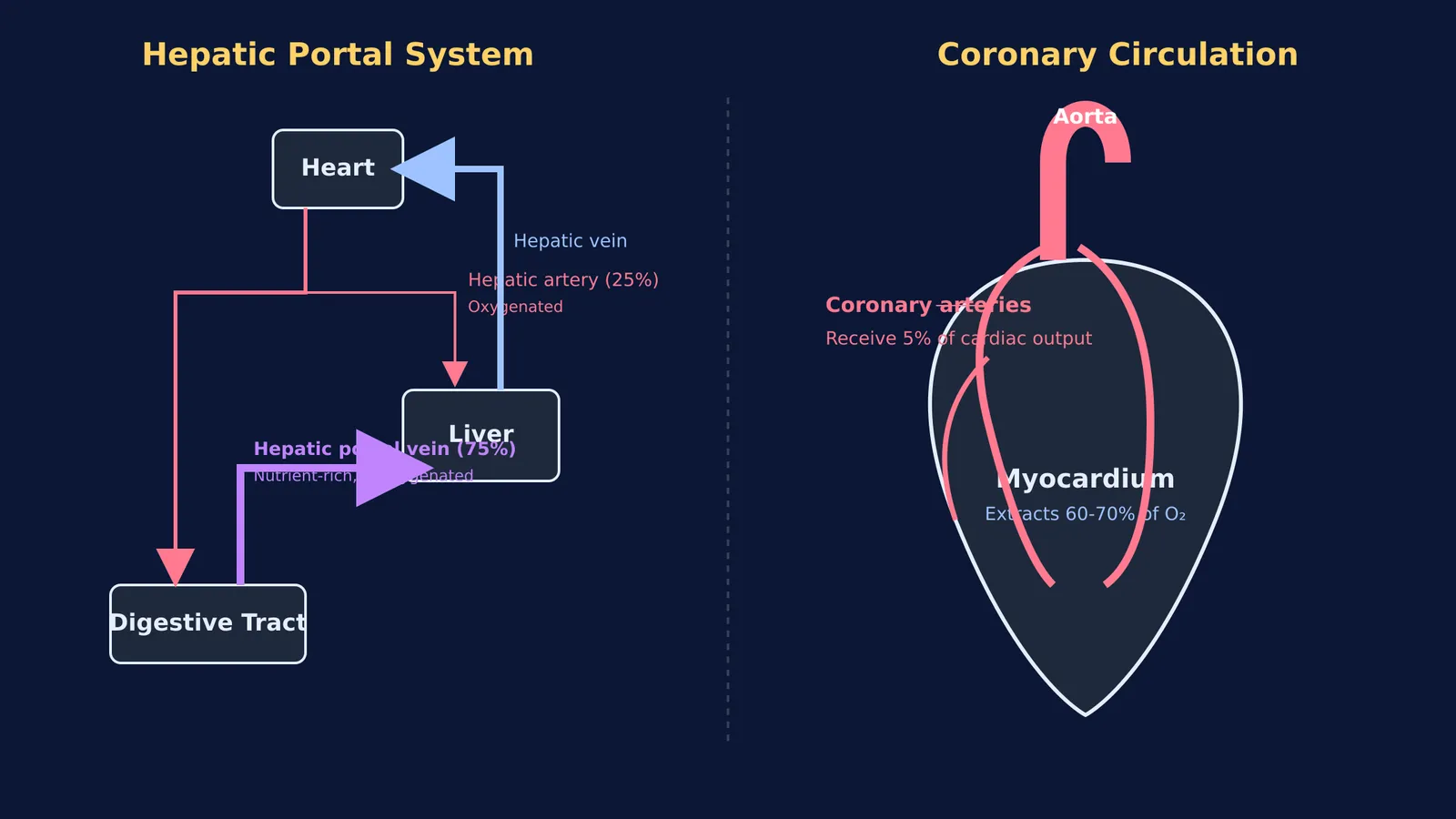
The hepatic portal system collects blood from your gastrointestinal tract—including the stomach, small and large intestines, pancreas, and spleen—and routes it directly into the liver. This venous blood is packed with raw nutrients absorbed from your recent meal, alongside formed elements of blood and potential toxins that require immediate filtering.
The hepatic portal vein provides roughly 75% of the liver's total blood flow (NCBI StatPearls). The remaining 25% is delivered by the hepatic artery, which supplies the highly oxygenated blood that the liver tissue needs to survive and function.
Why does the liver need a portal vein?
The liver acts as your body's primary biochemical shield and metabolic processing plant. If nutrient-rich blood from the intestines went directly to the heart and was pumped systemically, your blood sugar and nutrient levels would spike wildly after every meal, creating a dangerous physiological imbalance.
By routing this blood through the hepatic portal system first, the liver is given the opportunity to store excess glucose as glycogen, process incoming amino acids, and neutralise harmful substances before they can reach delicate organs like the brain. Once the liver has processed these materials within its microscopic capillary beds (called sinusoids), the blood finally exits via the hepatic veins. It then joins the inferior vena cava to return safely to the heart.
How does coronary circulation work?
While the heart is uniquely responsible for pumping blood to the entire body, the blood flowing directly through its internal chambers does not nourish the heart muscle itself. The myocardium (the muscular wall of the heart) is far too thick for oxygen to simply diffuse across the chamber walls and into the tissue.
Instead, the heart relies on its own dedicated and highly efficient network of blood vessels. The left and right coronary arteries branch off directly from the base of the aorta, ensuring the heart is the very first organ to receive freshly oxygenated blood immediately after it leaves the left ventricle. This supply must keep up with the rhythm set by the heart's electrical system, including the atrioventricular node.
The coronary circulation receives about 5% of the total cardiac output at rest (NCBI StatPearls). Despite this relatively small initial volume, the heart is incredibly efficient at using the resources it gets. The heart muscle extracts between 60% and 70% of the oxygen from the blood it receives, a much higher fraction than most other tissues (NCBI StatPearls).
When the coronary arteries become narrowed or blocked by plaque, the myocardium is deprived of oxygen, leading to a condition known as ischemia, which can ultimately result in a heart attack.
When you study these intricate networks, mapping them out visually is often the most effective strategy. If you are struggling to trace how these vessels connect, the Fermi Concept Library has every concept in biology, each with its own lesson and its place in the map, to help you build strong mental models of these mechanisms.
How can you study complex circulatory pathways?
Memorising these pathways from a static textbook diagram can feel overwhelming. To make the learning process more active and retain the information for your exams, try this three-step tracing framework:
Identify the source: Where exactly does the blood in this specific vessel come from?
Note the payload: Is the blood currently rich in oxygen, carbon dioxide, or recently digested nutrients?
Trace the destination: Which organ is receiving this blood next, and what physiological purpose does that connection serve?
Using this framework, try to sketch the primary pathways on a blank piece of paper without looking at your notes. Drawing the anatomical structures physically forces your brain to build stronger spatial relationships, moving your understanding from short-term memorisation into deep, durable comprehension.


