On this page
Katherine Esau studied the inside of plants for more than seven decades, and one tissue kept drawing her back: the phloem, which carries food. Ray F. Evert's biographical memoir of Esau for the US National Academy of Sciences says that throughout her career she kept researching phloem in two ways: how phloem-limited viruses affect plant structure and development, and how the sieve tube is built as a conduit for food. If you are preparing for NEET, you have met her already. NCERT's Class 11 Biology opens Unit 2 with a box about her (NCERT Class 11 Biology, Unit 2).
Who was Katherine Esau?
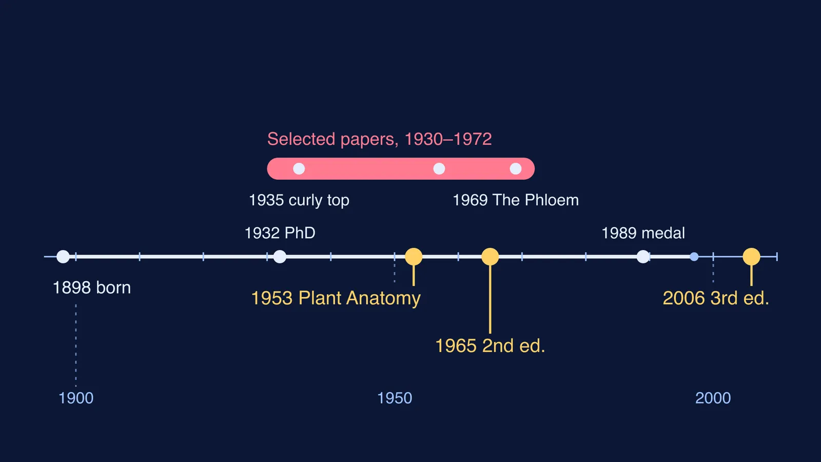
According to the NAS memoir, Esau was born on 3 April 1898 in Yekaterinoslav, in present-day Ukraine. Her Mennonite family fled to Germany at the end of 1918 and left for the United States in October 1922. In California she bred sugar beets to resist curly-top disease at the Spreckels Sugar Company. She arrived at the University of California, Davis, in 1927 and earned her PhD from Berkeley in 1932. She moved to UC Santa Barbara in 1963 and died on 4 June 1997, aged 99.
How did Katherine Esau link viruses to the phloem?
At Davis, Esau first planned to breed a beet that could resist the curly-top virus. To test it, she would have had to release the beet leafhopper, the insect that carries the virus, into university fields. Other researchers and growers objected. So she studied how the virus is transmitted and what it does to the sugar beet's phloem. In the memoir's words, she turned "from applied to the more basic study of plant anatomy as it relates to the disease" (NAS memoir).
Her early papers reported that the curly-top virus spreads through a plant by way of the food-conducting tissue, the phloem (NCERT Class 11 Biology, Unit 2). The memoir's selected bibliography lists a 1957 paper on phloem degeneration in grasses infected by barley yellow dwarf virus, and in 1967 she wrote the review Anatomy of Plant Virus Infections for the Annual Review of Phytopathology. She was introduced to electron microscopy just before she moved to Santa Barbara and applied it to her research there. The electron microscope facility now bears her name (NAS memoir).
For a closer look at the cells, see our guide to sieve tubes and companion cells. The story of Ernst Münch and the pressure flow hypothesis explains how food moves through them.
Why was Plant Anatomy a standard textbook for decades?
Plant Anatomy appeared in 1953, and the NAS memoir says it was known worldwide as "the bible of plant anatomy". Anatomy of Seed Plants followed in 1960. Both books were published in several languages, including Russian. A second edition of Plant Anatomy came out in 1965, and her book The Phloem (1969) reviewed the tissue from its earliest records.
The book has outlived her. Wiley published a third edition, Esau's Plant Anatomy, in 2006, by Ray F. Evert and Susan E. Eichhorn. Wiley's page for the book quotes a 2008 review in the journal Taxon, which says the 1953 first edition "became an instant classic and ushered in a new era in plant anatomy." Her 1989 National Medal of Science citation praised her "superlative performance as an educator, in the classroom and through her books" (NAS memoir).
Which Esau dates should you remember for exams?
The NCERT box and the memoir don't agree on every date. NCERT gives 1931 for her doctorate and 1954 for Plant Anatomy (NCERT Class 11 Biology, Unit 2). The NAS memoir gives 1932 and 1953, and Wiley's page for the book also gives 1953.
How do you find the phloem in a vascular bundle?
NCERT's Anatomy of Flowering Plants chapter describes the kinds of vascular bundle in a way you can turn into four questions for any transverse section:
Where are the xylem and the phloem? Together they form the vascular bundles.
Do they lie on alternate radii? That arrangement is called radial, as in roots.
Do they lie on the same radius? That is conjoint, common in stems and leaves, and the phloem usually sits on the outer side of the xylem.
Is there cambium between them? With cambium (dicot stems) the bundle is open. Without it (monocots) the bundle is closed.
Worked example: take the young dicot stem in NCERT Figure 6.4a. The phloem lies outside the xylem on the same radius, so the bundle is conjoint. A cambium layer lies between them, so it is open. Answer: conjoint and open. Now try the dicot root in Figure 6.3a. The xylem and phloem patches alternate, so the arrangement is radial.
If open, closed and conjoint still blur together, the Fermi Concept Library gives every biology concept its own lesson and its place in the map. For more practice, see our guide to identifying any T.S. in the anatomy of flowering plants.
Next time you draw a T.S. of a stem, find the phloem first, then work out what kind of bundle you are looking at.