On this page
When you lift a pen, protein threads in your muscles slide past one another. Before 1954 most scientists thought muscle shortened because its proteins folded up. Jean Hanson, a British zoologist, helped change that with Hugh Huxley by measuring the stripes in muscle and showing that one of them kept its length.
Who was Jean Hanson?
Emmeline Jean Hanson was born on 14 November 1919 and died suddenly on 10 August 1973, "at the height of her career", according to her Royal Society biographical memoir by John Randall. A historical review by the biochemist Koscak Maruyama in the Journal of Biochemistry gives the rest of the story. She was born in Newhall, Derbyshire, and graduated in zoology from Bedford College, London, in 1941. In 1947 she joined Randall's Medical Research Council biophysics unit at King's College London.
At King's, Hanson began studying isolated myofibrils, the thin strands inside a muscle fibre, under a phase-contrast microscope. Maruyama reports that she saw glycerinated rabbit myofibrils contract slowly to about 57% of their original length when ATP was added. In February 1953 a Rockefeller Foundation fellowship took her to the Massachusetts Institute of Technology (MIT).
How did Jean Hanson and Hugh Huxley discover the sliding filament theory?
At MIT she found Hugh Huxley, a young physicist whose electron micrographs had shown a double array of filaments in muscle. They combined her light microscopy with his electron microscopy.
First, they soaked muscle fibres in a solution that dissolves myosin, and the dark A bands largely disappeared. That showed the thick filaments of the A band are made of myosin, and strongly suggested that the thin filaments left behind were actin. They reported this in Nature in 1953.
In their 1954 Nature paper, they tracked the bands as myofibrils contracted and were stretched. Maruyama summarises what they found:
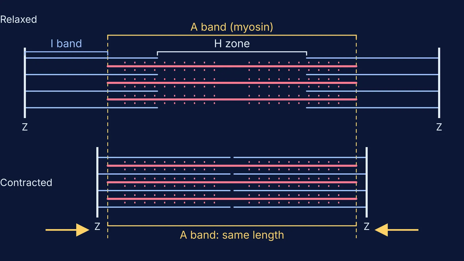
The A band stayed the same length as the sarcomere shortened, down to about 65% of its resting length.
The change in sarcomere length was taken up by the I band alone.
The H zone in the middle of the A band closed up during contraction.
When the myofibrils were stretched, the I band and H zone both got longer.
The simplest explanation was that two sets of filaments, each of fixed length, slide past one another. Andrew Huxley and Rolf Niedergerke, working separately in Cambridge, reached a similar conclusion using an interference microscope on living frog muscle fibres. The two papers appeared in the same issue of Nature on 22 May 1954.
Why did scientists doubt the sliding filament theory at first?
Maruyama explains that the prevailing view, shaped by the biophysicist William Astbury, was that muscle contracted through changes in the shape of its protein filaments. When Hanson spoke at a symposium in Leeds in September 1954, Astbury and other physicists were negative about the idea. Maruyama, writing in 1995, recalled her shouting: "I know I cannot explain the mechanism yet, but the sliding is a fact."
The idea was gradually accepted by the mid-1960s. Andrew Huxley proposed a cross-bridge theory in 1957 to explain what drives the sliding, and in 1963 Hanson and Jack Lowy showed that the actin filament is double-stranded.
How do you use this in a Class 11 answer?
The NCERT Class 11 Biology chapter on Locomotion and Movement says that during contraction "the 'I' bands get reduced, whereas the 'A' bands retain the length". That sentence is Hanson and Huxley's 1954 result.
Suppose a sarcomere (Z line to Z line) is 2.5 μm long at rest, and its A band is 1.5 μm, the width given for mammalian muscle in Maruyama's review. The 2.5 μm figure is for illustration only.
At rest: I band total = 2.5 − 1.5 = 1.0 μm, split as 0.5 μm on each side of the A band.
Contract to 65%: the new sarcomere is 0.65 × 2.5 = 1.625 μm.
The A band is still 1.5 μm, so the I band total is now 1.625 − 1.5 = 0.125 μm.
The H zone shrinks too, because actin from both ends now reaches further into the A band.
Check any answer on muscle contraction against this list:
A band: same length. Myosin filaments do not shorten.
I band: shorter. Actin overlaps myosin more.
H zone: shorter, or gone at full contraction.
Sarcomere: shorter, because the Z lines are pulled closer together.
Filaments: none of them change length. Writing that actin or myosin shortens is a common slip.
Related guides cover which sarcomere bands shorten during contraction, the skeletal muscle fascicle and sarcolemma and striated, smooth and cardiac muscle.
If sliding bands are hard to picture, the Fermi Simulation Lab has interactive simulations for ideas that are easier to see than to read. If you mix up actin, myosin and troponin, the Fermi Concept Library gives each concept its own lesson.
Next time you look at Figure 17.5 in your NCERT textbook, cover the labels and find the one band that stays the same. That is where Jean Hanson started.