

On this page
Understanding how a skeletal muscle contracts requires zooming in through several layers of anatomy. What looks like a single solid muscle is actually a bundle of bundles, wrapped in connective tissue and filled with specialised fluids and calcium stores. This "Russian doll" structure ensures that the electrical signals from your nervous system reach every contractile protein almost instantly.
What is the structure of a skeletal muscle fascicle?
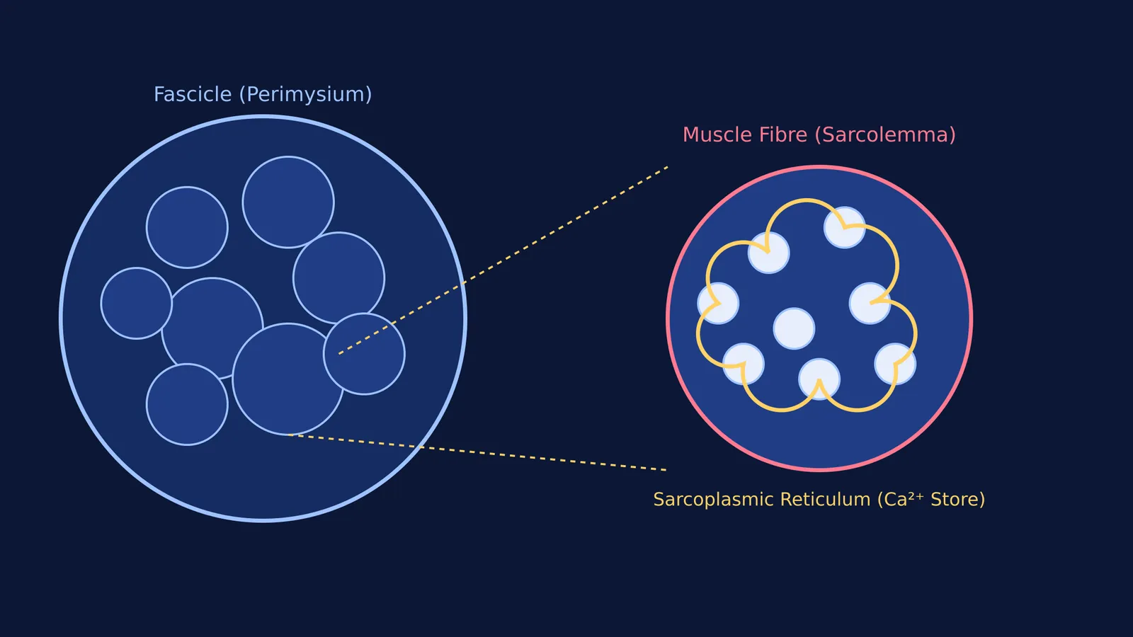
If you cut through a skeletal muscle, you will see it is divided into discrete bundles. Each bundle is a fascicle. The fascicles are held together by a middle layer of connective tissue called the perimysium. Zooming out, all of these fascicles are bundled together to form the complete skeletal muscle, which is encased in a tough, outer fibrous sheath known as the epimysium. This continuous network of connective tissue extends beyond the muscle to form the tendons that attach muscle to bone, ensuring that when the muscle shortens, the force is transferred effectively to move the skeleton.
Inside the fascicle lie the individual muscle cells, which are known as muscle fibres. Because they are built to generate force, these fibres are remarkably long and cylindrical, with a typical diameter ranging from 10 to 100 micrometers (µm) (NCBI Bookshelf: Physiology, Skeletal Muscle). Every single fibre is wrapped in its own delicate connective tissue sheath called the endomysium. This separation allows the nervous system to recruit specific fascicles or fibres for a task, rather than firing the entire muscle at once.
If you keep confusing the epimysium, perimysium and endomysium, Fermi's AI tutor can build a plan to review the connective tissue layers and check your understanding as it goes.
What is the sarcolemma and the sarcoplasm?
Just beneath the endomysium lies the muscle fibre's true outer boundary: the sarcolemma. This is the specialised plasma membrane of the cell. Like a standard cell membrane, it is a phospholipid bilayer, but it is adapted to carry electrical action potentials at a speed of 3 to 5 metres per second (Physiology, Skeletal Muscle).
To ensure that the electrical signal does not just skim the surface of the cell, the sarcolemma plunges inward to form transverse tubules (T-tubules). These tiny tunnels carry the action potential deep into the core of the fibre.
Inside the sarcolemma is the sarcoplasm, which is the muscle fibre's equivalent of cytoplasm. It is densely packed with mitochondria, which supply the massive amounts of ATP required for muscle activity, along with glycogen granules that serve as an immediate energy reserve. The sarcoplasm is also filled with hundreds or even thousands of rod-like myofibrils. These myofibrils are the actual contractile machinery of the cell, composed of repeating units called sarcomeres that contain perfectly overlapping threads of thick myosin and thin actin filaments.
How does the sarcoplasmic reticulum act as a calcium store?
Woven tightly around the myofibrils in the sarcoplasm is the sarcoplasmic reticulum (SR). This is a specialised smooth endoplasmic reticulum whose main job is to store and release calcium ions (Ca²⁺).
The SR maintains an enormous concentration gradient. At rest, the SR stores calcium at a concentration approximately 10,000 times higher than the free calcium circulating in the sarcoplasm (Physiology, Skeletal Muscle). To manage this vast stockpile, the SR contains a protein called calsequestrin, which binds up to 40 calcium ions per molecule.
The structure of the SR aligns perfectly with the T-tubules to form a triad: one T-tubule flanked by two enlarged sacs of the SR called terminal cisternae.
When an action potential shoots down the T-tubule, voltage sensors in its membrane (known as dihydropyridine receptors) change shape. They mechanically pull open the ryanodine receptors on the adjacent SR membrane, immediately flooding the sarcoplasm with calcium. The sudden spike in calcium uncovers the binding sites on the actin filaments by moving the regulatory proteins troponin and tropomyosin out of the way. This allows the myosin heads to grab the actin and pivot, pulling the filaments past one another and allowing the entire muscle to contract forcefully.
When you are trying to picture how T-tubules and the terminal cisternae fit together in 3D, static textbook diagrams can be confusing. This is where the Fermi Concept Library helps, giving the molecular machinery of contraction its own lesson and its place in the map.
Once the signal stops, calcium pumps (SERCA) in the SR membrane burn ATP to rapidly drag the calcium back inside, allowing the muscle to relax and prepare for the next beat.
If you are studying the wider systems that support these muscle movements, you can read our guides to the human skeleton: counting axial and appendicular bones and August Krogh and how capillaries open when muscles work.


