On this page
In the early spring of 1958, David Hubel met Stephen Kuffler and Torsten Wiesel in a cafeteria at Johns Hopkins Hospital in Baltimore, as Hubel recalled in his 1981 Nobel lecture. Hubel was born in Windsor, Canada, in 1926, and Wiesel in Uppsala, Sweden, in 1924 (Nobel Prize facts pages for Hubel and Wiesel). Within a week of Hubel arriving at Hopkins, the pair were recording single cells in the visual cortex of anaesthetised cats. What they found: the brain breaks an image into short edges, each handled by cells tuned to one angle.
What did Hubel and Wiesel discover?
For about a month, the cells ignored the spots of light the pair shone onto the cat's retina. Then, during one recording that lasted 9 hours, they slid a glass slide with a black dot into the slot of their ophthalmoscope and the cell "went off like a machine gun", Hubel wrote in the Nobel lecture. The edge of the glass was casting a faint, sharp shadow on the retina: a straight dark line on a light background. In Hubel's words: "That was what the cell wanted, and it wanted it, moreover, in just one narrow range of orientations."
Their first full paper, Receptive fields of single neurones in the cat's striate cortex, appeared in The Journal of Physiology in October 1959. A longer 1962 paper in the same journal, on receptive fields, binocular interaction and functional architecture, mapped how these cells are organised and how input from the two eyes combines (see why we need two eyes to judge depth).
Why do visual cortex neurons respond to lines and not spots?
A receptive field is the patch of the retina where light can change a particular cell's firing. Hubel's Nobel lecture describes the first type they understood, which they called simple cells.
Simple cells have receptive fields split into "on" and "off" strips separated by parallel straight lines, set vertical, horizontal or oblique.
Light over the whole field gives almost no response, because the excitation and inhibition cancel "with the precision of an acid-base titration".
A line turned 90° from the best angle has no effect wherever it is placed.
Complex cells respond to a line at the right angle anywhere in their field, fire hardest when it moves, and about half prefer one direction of movement.
Their explanation was a ladder: a complex cell may collect input from many simple cells that share one orientation. Hubel cautioned that these cells are no more "line detectors" than curve detectors, and that how the brain assembles them into what we perceive was "still a complete mystery".
How are orientation cells arranged in the cortex?
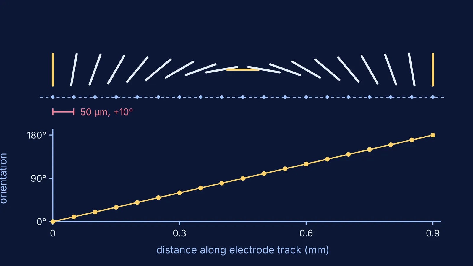
In 1961, in a spider monkey, they pushed an electrode 2.5 mm through the cortex at a slant, Hubel recalled in the Nobel lecture. The preferred orientation moved in small steps of about 10° for every 50 µm the electrode advanced. In five hours they logged 53 orientations in a row.
The Nobel Prize press release sums it up: the cells are arranged in columns, cells within a column share the same function, and these group into hypercolumns, each covering about 2 by 2 millimetres of cortex. For the older map of the cortex, see Korbinian Brodmann and the 52 areas of the cerebral cortex.
Why does early visual experience matter?
The same press release says Hubel and Wiesel showed that this ability develops directly after birth, and that light alone is not enough: the image must have pattern and many contours. Dull or distorted images in early life, for example from errors in the eye's lens system, may permanently impair how the brain analyses what it sees.
The image these cells read starts upside down on the retina: see how Kepler proved the eye sees the world upside down.
Try it: predict what one cortex cell will do
Take a simple cell whose best stimulus is a vertical bright slit on its "on" strip. Cover the answers and predict each response.
The whole screen lights up evenly: almost no response, because on and off regions cancel.
A vertical slit lies exactly on the on strip: strong firing.
The same slit shifts sideways to a parallel position over an off strip: firing drops sharply.
The slit is turned horizontal: no response, wherever it sits.
A complex cell tuned to vertical, with a vertical line swept sideways anywhere in its field: strong firing that fades soon after the line stops.
A white egg on a dark background: Hubel's own example. Cells whose fields sit inside the egg stay quiet; only cells cut by its border, where the border's angle matches theirs, fire.
If the neuron or visual pathway behind this is still hazy, the Fermi Concept Library has a lesson for every biology concept with its place in the map. Fermi's AI tutor can then work through a list like this with you, deciding whether to explain, ask or show, and checking your understanding as it goes.
Next time you read a page, picture cells each watching for one short edge at one angle.