On this page
If you look at the folded surface of the human brain, it appears mostly uniform. However, beneath that smooth surface lies a highly specialised division of labour. The map we use to navigate this territory was drawn over a century ago by a German neurologist named Korbinian Brodmann, who chose to look at the brain not by what it did, but by how it was built.
How did Brodmann map the cerebral cortex?
In the early 20th century, the scientific community was fiercely debating whether the brain operated as a single, indivisible organ or if specific functions were localised to distinct areas. In 1901, Brodmann joined the Neurobiological Laboratory in Berlin, working alongside the husband-and-wife research team of Oskar and Cécile Vogt.
While the Vogts focused their research on myeloarchitectonics (the study of myelinated nerve fibres), they tasked Brodmann with studying cytoarchitectonics—the microscopic organisation, density, and types of cell bodies within the brain tissue.
Using the Nissl staining method, a technique that selectively dyes the cell bodies of neurons, Brodmann examined extremely thin slices of the cortex under a microscope. The cerebral cortex is typically made up of six layers of cells (Source). As Brodmann moved his microscope across different regions of the brain, he noticed that these layers changed noticeably in their thickness, cell size, and cell types.
By tracing these microscopic boundaries, he divided the human cerebral cortex into 52 distinct areas. He published his detailed, numbered map in his 1909 monograph *Vergleichende Lokalisationslehre der Großhirnrinde* (Comparative Localization in the Cerebral Cortex) (Source).
This structural approach was a massive task of observation. If you are learning the broader anatomy that these areas sit within, our guide to the Parts of the human brain: forebrain, midbrain and hindbrain roles covers the wider geography.
What are the most common Brodmann areas?
Brodmann did not know the function of every area he mapped, but he operated on the principle that a change in cellular structure indicates a change in function. This assumption proved remarkably accurate. The structural map he created mapped closely onto functional roles that were discovered later.
While modern techniques have identified further subdivisions, Brodmann's original numbering remains the vocabulary of neuroanatomy. Here are the most frequently referenced areas:
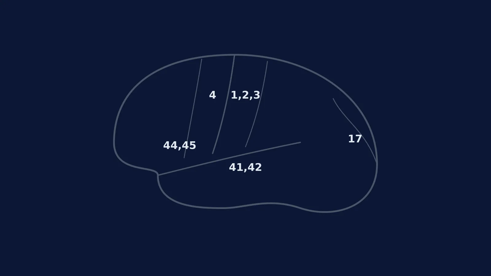
Areas 1, 2, and 3: The primary somatosensory cortex, located just behind the central sulcus. This region processes sensory information like touch, temperature, and pain.
Area 4: The primary motor cortex, located just in front of the central sulcus. It is responsible for the execution of voluntary muscle movements.
Area 17: The primary visual cortex, situated at the very back of the brain in the occipital lobe. It processes basic visual stimuli.
Areas 41 and 42: The primary auditory cortex, located in the temporal lobe, responsible for processing sound.
Areas 44 and 45: Known as Broca's area (typically in the left hemisphere), this region is crucial for speech production and language processing.
Why do we still use Brodmann areas today?
Despite being drawn from two-dimensional histological slices over a century ago, Brodmann's 52 regions remain the standard coordinate system in modern neuroscience (Source).
When a patient undergoes a functional magnetic resonance imaging (fMRI) or PET scan to see which part of their brain is active during a cognitive task, researchers still report the active locations using Brodmann's numbers. The cellular arrangement of the primary motor cortex (Area 4) looks distinctly different from that of the primary visual cortex (Area 17), and those structural boundaries align closely with the functional boundaries we can now observe in living, thinking brains.
The Vogts were highly ambitious and eventually proposed a map of over 200 distinct areas based on their nerve fibre research (Source), but Brodmann's simpler, 52-area map proved more resilient. Sadly, Brodmann did not live to see the full impact of his work; he died suddenly from a severe infection in 1918 at the age of 49 (Source). However, his map quickly became the universal language of brain research, especially after it was adopted by neurosurgeons to localise brain injuries.
As other pioneers developed new ways to study the nervous system in the decades that followed—such as Wilder Penfield and mapping the brain during surgery—they continually validated the functional borders that Brodmann had predicted from cell structure alone.
When you are studying these regions, keeping track of how they connect can be challenging. The Fermi Concept Library has every concept in physics, chemistry, biology, mathematics and computing, each with its own lesson and its place in the map, so you can trace how a specific cortical area relates to the rest of the nervous system.