On this page
Why were nerves confused with tendons before Herophilus?
For centuries, Greek natural philosophy operated without opening a human cadaver. Religious customs, civic laws, and anxieties surrounding corpses strictly forbade dissecting human bodies across the Aegean world. As classical scholar Heinrich von Staden documented in The Yale Journal of Biology and Medicine, touching or cutting human flesh after death was viewed as severe religious contamination.
Deprived of internal direct observation, early Greek thinkers relied on animal butchery and speculation. In archaic Greek, the word *neuron* described any tough, fibrous cord, including a bowstring, a lyre string, or a sinew. When Aristotle (384–322 BC) examined animals, he observed fibrous strands near joints and concluded that all bodily movement was directed by the heart. As neurobiologist Eugenio Frixione explained in the Journal of the History of the Neurosciences, Aristotle grouped peripheral nerves, tendons, and ligaments together under this single concept of *neura*. In Aristotle's cardiocentric model, the heart served as the central furnace of sensation, while the brain was merely a moist organ that cooled circulating blood. Tendons that pulled bones and cords that carried sensation were lumped together.
How did Herophilus prove nerves originate in the brain?
That confusion ended around 300 BC in Alexandria. Under Ptolemy I Soter and Ptolemy II Philadelphus, traditional taboos were temporarily set aside. For an unprecedented window of roughly 30 to 40 years, anatomists were permitted to systematically dissect human cadavers, as medical historians Noel Si-Yang Bay and Boon-Huat Bay noted in Anatomy & Cell Biology.
Herophilus of Chalcedon (c. 335–255 BC) seized this opportunity. Rather than relying on philosophical speculation, he placed a scalpel into real human tissue, carefully tracing the body's white cords along their full paths.
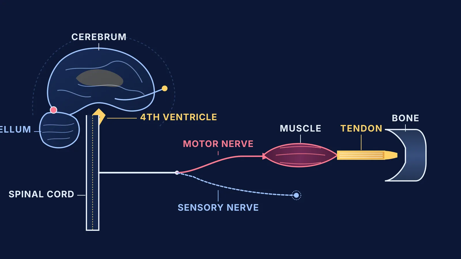
Herophilus observed that fibrous cords were not identical in origin or destination. While tendons originated in muscular bellies and inserted into bone, other white fibres could be followed inwards through the skull and vertebrae. As neurologist J. M. S. Pearce detailed in European Neurology, Herophilus traced these true nerves directly back to the cerebrum, cerebellum, and spinal cord, proving they did not originate in the heart.
Furthermore, Herophilus recognised a functional divide between two classes of nerves: sensory nerves conveying perception, and motor nerves (*neura proairetika*) transmitting impulses for voluntary movement. Through careful observation, he noted that damaging these motor nerves caused paralysis, even when muscles and tendons remained structurally intact.
What anatomical structures did Herophilus discover?
Herophilus did not stop at peripheral nerve pathways; he systematically charted the human cranium. His detailed mapping laid the foundations of neuroscience, a milestone analysed by Adrian Wills in The Lancet02081-4).
Inside the skull, Herophilus identified and named key structures still taught today:
Cranial nerves: He identified seven pairs of cranial nerves, including the optic nerve for vision, the oculomotor nerve for eye movement, and the trigeminal and hypoglossal nerves.
Cerebral ventricles: He mapped the internal chambers of the brain, identifying the fourth ventricle as the primary centre of motor coordination, and named the *calamus scriptorius* after its resemblance to a reed pen.
Meninges and venous sinuses: He described the protective membranes enveloping the brain and mapped the confluence of cranial venous sinuses, later named the *torcular Herophili*.
The eye: He dissected the eye's coats, differentiating the cornea, retina (which he described as a casting net), iris, and choroid layer.
Although his written treatises were lost during antiquity, his empirical breakthroughs survived through later Roman and Islamic commentaries. Centuries later, Galen's anatomical experiments and errors relied on Herophilus's baseline descriptions, and during the Renaissance, Andreas Vesalius and the revival of human dissection returned directly to Herophilus's standard of verifying anatomy through direct human inspection.
How can students separate neural control from mechanical motion?
For students mastering the parts and functions of the human brain, confusing neural signalling with musculoskeletal motion remains a frequent conceptual pitfall. In examinations like NEET-UG, questions regularly test whether candidates understand the functional divide between tissue types and reflex pathways.
Here is a 4-step framework to distinguish neural from musculoskeletal tissue:
Trace anatomical origin: True nerves originate from the neuroaxis (brain or spinal cord), whereas tendons emerge from muscle bellies and insert into bone periosteum.
Identify tissue architecture: Nerves consist of bundled axons enclosed in perineurium and myelin sheaths, while tendons consist of dense regular connective tissue packed with parallel collagen fibres.
Determine functional transmission: Nerves propagate electrochemical action potentials for sensory input and motor commands, whereas tendons passively transmit mechanical force.
Predict lesion outcomes: Severing a motor nerve causes flaccid paralysis without tendon damage, while a ruptured tendon leaves neural firing intact but destroys joint actuation.
When studying human physiology, students often struggle to separate mechanical linkages from electrical signalling networks. The Fermi Concept Library covers every concept in physics, chemistry, biology, mathematics and computing, each with its own lesson and its place in the map. For students tackling neuromuscular coordination problems, the Fermi Problem Library provides worked problems with a step-by-step solver that explains the reasoning rather than printing an answer.