On this page
When the anatomist Gaspare Aselli opened a well-fed dog in Milan in 1622, he found fine white vessels running through the membrane that holds the intestine. They were the lacteals, the lymph vessels that carry fat away from your gut, and his account is where the first true description of the lymphatic system begins. Here is what he saw, why the meal mattered, and where it fits your syllabus.
Who was Gaspare Aselli and what did he see in 1622?
Aselli was an anatomist and surgeon from Cremona who worked in Milan and took the chair of anatomy at Pavia in 1624, according to a 2023 history of the lymphatic system by surgeon Giancarlo Pansini in Veins and Lymphatics.
On 23 July 1622, urged by friends, Aselli cut open a living dog that had been fed, planning to show its recurrent nerves and the movement of its diaphragm (Pansini, 2023). Pulling the intestines aside, he noticed, in the translation Pansini quotes, "numerous white cords, exceedingly thin and beautifully white, branched throughout the mesentery and on the peritoneal surface of the intestine". At first they could have passed for nerves. Then he cut one, and a whitish fluid like milk or cream ran out.
Why did the white vessels appear only in a fed dog?
The next day Aselli opened a second dog and could not find the vessels (Pansini, 2023). He suspected the reason: this dog had been opened on an empty stomach. He then opened a third dog that had eaten a few hours earlier, and the white vessels were back. After seeing them in other mammals, he proposed the name vasa lactea, the milky vessels.
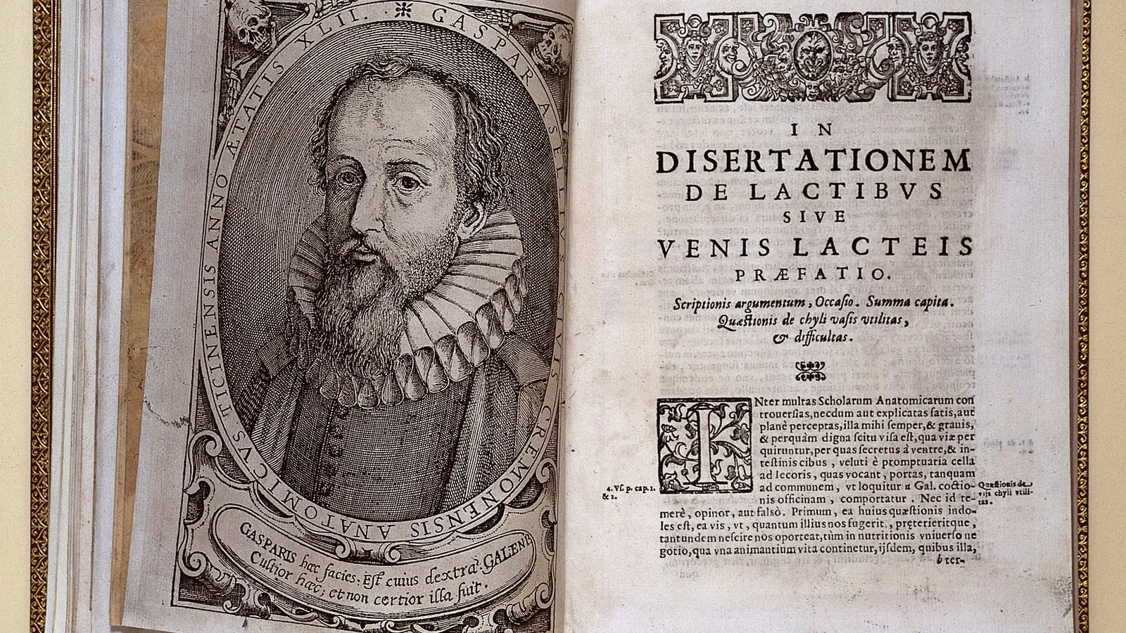
His book, De lactibus sive lacteis venis, was printed in Milan in 1627, after his death. Pansini notes that its four colour woodcuts were the first illustrations of lymphatic vessels and probably the first in colour in medical literature. Aselli still got one thing wrong: he believed the vessels carried their fluid to the liver, where it was supposedly turned into blood.
What do lacteals actually do with the fat you eat?
A lacteal is the lymph vessel in the central core of each villus, the finger-like projection lining the small intestine (Zhou, Qu, Liu and Tso, 2020).
Cells lining the intestine pack digested fat into large particles called chylomicrons, defined as at least 800 Å (80 nanometres) across, for export into the lymphatic system (Zhou et al., 2020). The same review credits Aselli with showing that particulate fat in a dog's mesenteric lymph rises after a meal. The milk he saw was fat.
The route is the part Aselli missed. Lacteals drain into the intestinal lymph duct, then the thoracic duct, and finally empty into the left subclavian vein (Zhou et al., 2020). The liver is not on that path. Pecquet described the thoracic duct in 1651, and in 1653 Bartholin introduced the term vasa lymphatica and stopped treating the liver as the centre of blood production (Pansini, 2023).
Where do lacteals fit in Class 10 and Class 11 biology?
The NCERT Class 10 chapter Life Processes says the villi increase the surface area for absorption and are "richly supplied with blood vessels which take the absorbed food to each and every cell of the body" (NCERT Class 10 Science, Chapter 5). It does not mention lacteals, so it is easy to assume fat follows the same path as sugar. It takes the lymph route first.
In CBSE's 2025-26 Class 11 Biology curriculum there is no separate digestion chapter: lipids appear in Biomolecules, and "composition of lymph and its function" sits in Body Fluids and Circulation (CBSE Biology curriculum 2025-26). Lacteals link those two chapters. For the lymph side, read our explainer on how lymph differs from plasma and tissue fluid.
How can you trace fat from your plate to your blood?
This five-stop trace joins the NCERT Class 10 text to the lymph route in Zhou et al.. Say each stop out loud, then draw it as one arrow chain.
Bile salts break large fat globules into smaller ones, so enzymes can act on them. Our post on how bile emulsifies fat for lipase explains the surface-area idea.
Lipase and intestinal enzymes break the fats down into fatty acids and glycerol.
Cells lining the intestine repack the digested fat into chylomicrons.
Chylomicrons enter the lacteal at the centre of the villus.
Lymph carries them through the thoracic duct into the left subclavian vein.
If a stop stays fuzzy, for instance what a lipoprotein is, the Fermi Concept Library gives every concept in biology, chemistry and physics its own lesson and its place in the map.
For another experiment that watched digestion inside a living body, read about William Beaumont and the stomach he could see into.