
On this page
In 1865 a 23-year-old Italian medical graduate, Enrico Sertoli, published a description of tall, branching cells inside the tubules of the human testis. You meet those cells today as Sertoli cells: the nurse cells that look after sperm while they develop. Here is who he was, what he saw, and a simple way to keep the facts straight for exams.
Who discovered Sertoli cells?
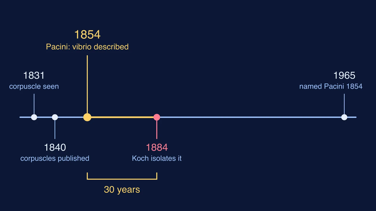
Enrico Sertoli was born in 1842 in Sondrio, in northern Italy, and died there in 1910, according to a 2019 historical article by Patriarca, Colecchia and Clerici in the journal Pathologica. He graduated in medicine from the University of Pavia in 1865, at 23.
The same year he published his description in the journal *Il Morgagni*, under the title "Dell'esistenza di particolari cellule ramificate nei canalicoli seminiferi del testicolo umano", which means "On the existence of special branched cells in the seminiferous tubules of the human testis" (Pathologica, 2019).
He went on to study in Vienna in the laboratory of the physiologist Ernst Brücke. In 1870, at 28, he was called to teach anatomy and physiology at the higher school of veterinary medicine in Milan, the same article records.
What did Sertoli actually see in 1865?
He saw cells shaped a little like trees. He called them cellule ramificate, branched cells, and also referred to them as cellule madri, mother cells, as a 2016 review in the journal Andrology recounts (França and colleagues, 2016).
The name came later and from someone else. In 1888 von Ebner first wrote of "Sertoli's cells", both the Andrology review and the Pathologica article note.
Sertoli kept working on the testis. In 1878 he published a study of the structure of seminiferous tubules and the development of spermatids in rats (Pathologica, 2019). Even so, the same article says, scientific attention to his cells "remained very limited for roughly half a century after Sertoli's death".
What do Sertoli cells do?
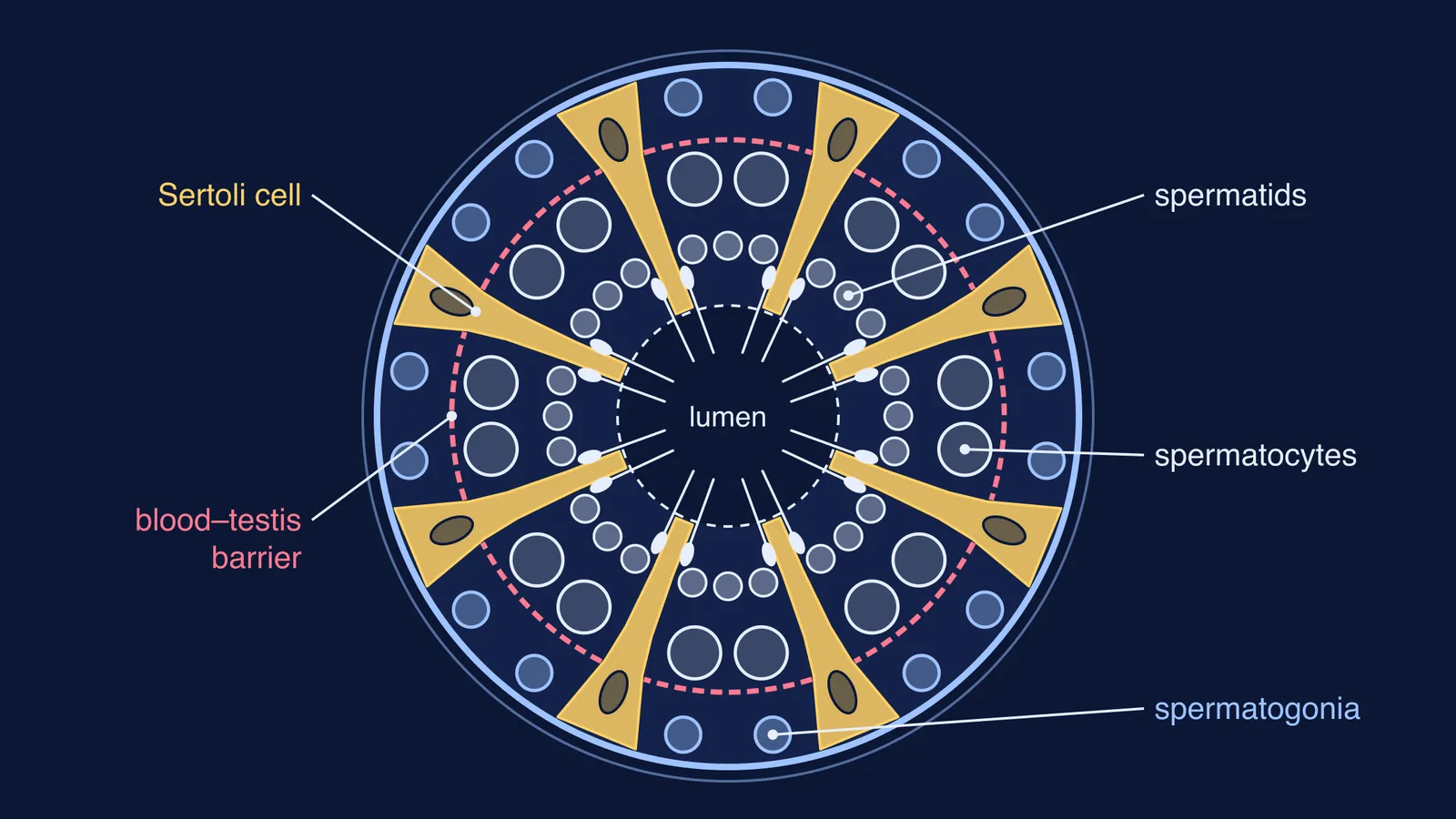
A Sertoli cell is a tall supporting cell that reaches from the outer wall of a seminiferous tubule to its lumen and surrounds the developing germ cells, the cells that become sperm (França and colleagues, 2016). The 2016 review lists jobs Sertoli could not have seen:
Feeding: Sertoli cells move micronutrients across the blood-testis barrier to support germ cell development.
Shelter: junctions between neighbouring Sertoli cells form the blood-testis barrier, which was defined physiologically in 1967 and seen under the electron microscope in 1970.
Clean-up: Sertoli cells engulf (phagocytose) germ cells that degenerate.
Hormone relay: they are the targets for FSH and testosterone and pass those signals on inside the testis.
Release: they move the most mature spermatids toward the lumen, where they are released in a step called spermiation.
The same review lists androgen-binding protein (1975) and inhibin (1979) among the products of Sertoli cells.
Each Sertoli cell can look after only so many developing sperm. The number of spermatids per Sertoli cell ranges from more than one hundred in fish to fewer than ten in most mammals studied, and is about four in humans (França and colleagues, 2016).
How do you tell Sertoli cells from Leydig cells?
Both cell types live in the testis, so they are easy to mix up. Leydig cells sit next to the seminiferous tubules rather than inside them, and they produce testosterone in the presence of luteinising hormone (LH); they are named after the German anatomist Franz Leydig, who identified them in 1850 (Leydig cell, Wikipedia).
Use this four-question recall card. Cover the answers and say them aloud.
Where is it? Sertoli: inside the tubule, from the outer wall to the lumen. Leydig: between the tubules.
Which pituitary hormone acts on it? Sertoli: FSH. Leydig: LH.
What does it make? Sertoli: androgen-binding protein and inhibin. Leydig: testosterone.
Who described it, and when? Sertoli: Enrico Sertoli, 1865. Leydig: Franz Leydig, 1850.
A memory hook: Sertoli and FSH both contain an S, and the S cell stays on the sperm side of the tubule wall. Leydig goes with LH.
How can you revise this so it sticks?
Draw it, do not just read it. Make a circle for the tubule, a smaller circle for the lumen, and a few tall columns from wall to lumen for the Sertoli cells. Then add, from the outside in, spermatogonia, the barrier line, spermatocytes and spermatids. Label it from memory, check it against your textbook, and explain each label aloud in your own words. The gaps show you what to reread.
If the hormone loop between the pituitary, Leydig cells and Sertoli cells still feels tangled, the Fermi Concept Library gives every concept in biology its own lesson and shows its place in the map, so you can see how spermatogenesis connects to hormones. And if you are not sure where to start with the reproduction chapter, Fermi's AI tutor works out what you already know and builds a plan for the session.
Next time you draw a seminiferous tubule, give the tall branched cells their proper credit: a 23-year-old graduate of Pavia described them in 1865.