On this page
Every sound you hear, from a teacher's voice to a pressure cooker whistle, must become an electrical signal before your brain can use it. That happens in a spiral strip of cells inside the cochlea. It is named after the young Italian anatomist who first described it in 1851, then left science within three years.
Who discovered the organ of Corti?
Alfonso Corti, an Italian anatomist born in 1822 in Gambarana, now in the province of Pavia, first described the organ of Corti, according to a 2023 biography by Mira, Martini, Mazzarello and Mudry in the Italian Journal of Audiology and Phoniatrics. He qualified in medicine in Vienna in 1847, then trained in anatomy labs across Europe, including Albert Kölliker's in Würzburg in 1850.
His paper, “Recherches sur l'organe de l'ouïe des mammifères” (research on the hearing organ of mammals), was printed on 30 June 1851 in the journal Zeitschrift für wissenschaftliche Zoologie. Kölliker quoted Corti's observations in his histology textbook and called the structure Corti's “spiral organ”, which helped the name spread.
What did Corti actually see?
The cochlea is soft tissue sealed inside the temporal bone, which made it very hard to study. Corti examined at least 200 cochleas from humans and other animals, as summarised in Eugenio Mira's 2023 paper on his life.
In about 60 pages, with two colour plates engraved from his own drawings, he described the cells of the inner ear, giving sizes and numbers with remarkable accuracy for his time (Mira and colleagues, 2023). In their translation, he wrote: “I am convinced that I am far from having exhausted such a difficult subject as the anatomy of the mammalian cochlea.”
He never got further. Dividing his father's inheritance pulled him back to Italy, and in 1854 he stopped scientific work completely. That year he also wrote of an illness that his biographers think was probably rheumatoid arthritis. He died in 1876.
How does the organ of Corti turn sound into nerve signals?
The US National Institute on Deafness and Other Communication Disorders (NIDCD) describes six steps:
Sound waves travel down the ear canal to the eardrum.
The eardrum vibrates, and the bones of the middle ear amplify the vibrations and pass them to the cochlea.
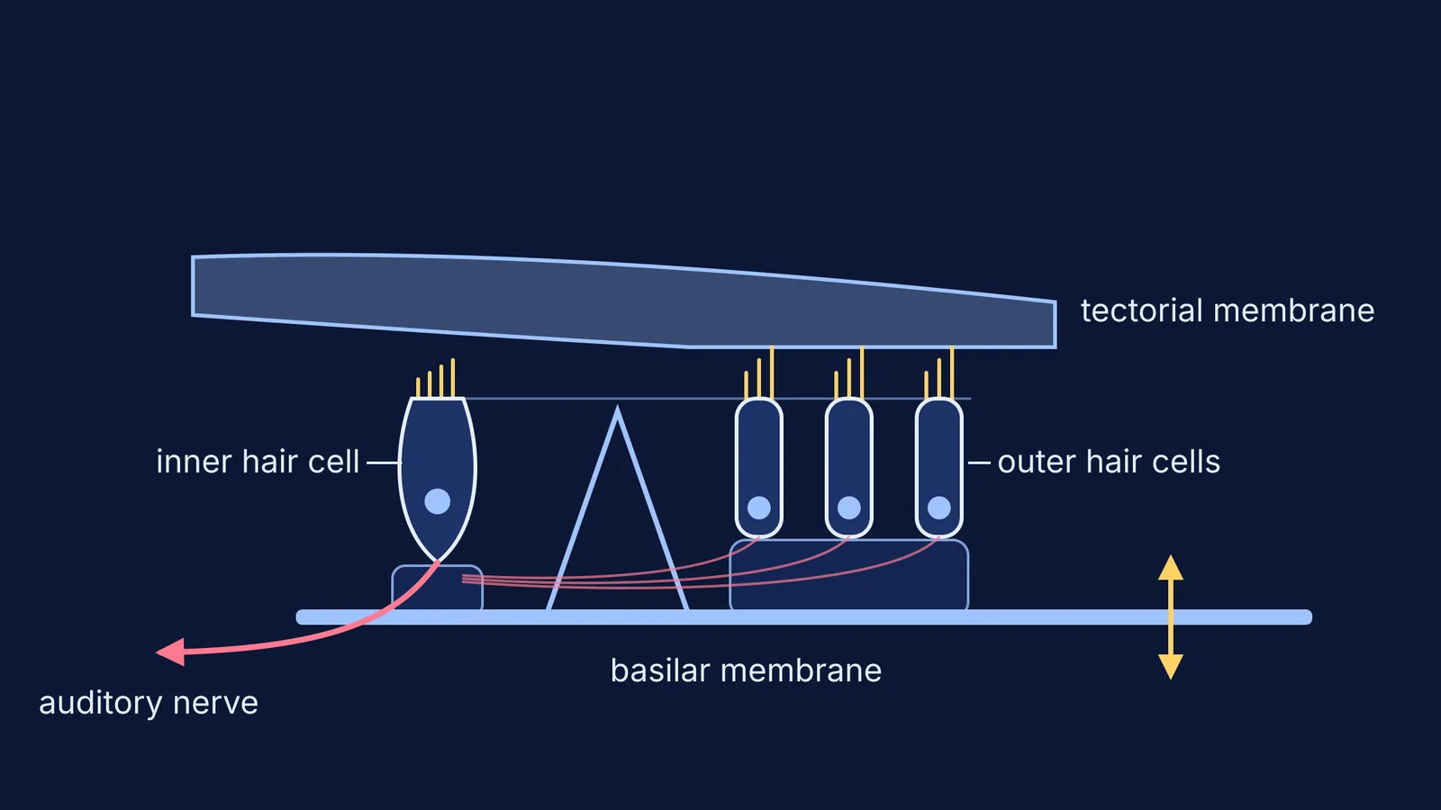
Inside the cochlea, a travelling wave forms along the basilar membrane, and the hair cells sitting on it move up and down.
Tiny projections on top of each hair cell, the stereocilia, bump against an overlying structure and bend.
Bending opens pore-like channels, chemicals rush into the cells, and an electrical signal is created.
The auditory nerve carries that signal to the brain.
That overlying structure is the tectorial membrane, in which the tips of the tall outer hair cell stereocilia are embedded (Avan and colleagues, PNAS 2019).
Each human cochlea has about 3,500 inner hair cells and about 11,000 outer hair cells, the outer ones in 3 or 4 rows (BioNumbers, citing Ashmore 2008). The inner hair cells are the main sensory cells. The outer hair cells act as tiny motors that amplify sound, driven by a protein called prestin (Ashmore, Physiological Reviews 2008).
Pitch has an address, too. Hair cells near the wide end of the snail-shaped cochlea detect high-pitched sounds and those closer to the centre detect low-pitched ones (NIDCD). Find your own high-pitch limit with our hearing range tone test.
If a wave travelling along a membrane is hard to picture, the Fermi Simulation Lab has interactive simulations for ideas that are easier to see than to read.
Why should you look after your hair cells?
Human hair cells do not grow back, and long or repeated exposure to sounds at or above 85 dBA can cause hearing loss, says the NIDCD page on noise-induced hearing loss. The World Health Organization estimates that over 1 billion young adults are at risk of permanent, avoidable hearing loss from unsafe listening (WHO fact sheet).
How do you remember the organ of Corti for exams?
Learn this 30-second answer:
The organ of Corti is the hearing organ inside the cochlea, first described by Alfonso Corti in 1851. It sits on the basilar membrane and carries one row of inner hair cells and three (sometimes four) rows of outer hair cells. When sound makes the membrane vibrate, the stereocilia bend, channels open, and the hair cells send electrical signals along the auditory nerve to the brain.
Then check yourself against these common mix-ups:
Location: the organ of Corti is in the inner ear, not among the middle-ear bones. Revise those with our guide to the ear ossicles: malleus, incus and stapes.
Rows: one row of inner hair cells, three (sometimes four) of outer hair cells.
Roles: inner hair cells sense; outer hair cells amplify.
Pitch map: high pitch at the wide end of the coil, low pitch towards the centre.
Match questions: Corti goes with the organ of hearing. Practise more pairs with our list of biology scientists and their discoveries.
If related ideas, such as how a nerve signal travels, feel shaky, the Fermi Concept Library gives each concept in biology its own lesson and its place in the map.