On this page
Before 1948, scientists knew that animal cells consumed oxygen and produced adenosine triphosphate (ATP) to store energy. They also knew that the enzymes for these reactions were attached to some insoluble structure in the cytoplasm. However, they did not know which specific organelle was responsible. The cell was still largely a black box.
In 1948, Albert Lehninger and Eugene Kennedy broke that box open. Using a new centrifuge technique to separate the parts of rat liver cells, they proved that the mitochondria contain the complete enzymatic machinery for oxidative phosphorylation.
Where is the site of oxidative phosphorylation?
The site of oxidative phosphorylation in eukaryotic cells is the inner membrane of the mitochondrion. Lehninger and Kennedy demonstrated this by isolating rat liver mitochondria and feeding them fatty acids and intermediates of the tricarboxylic acid (Krebs) cycle (Kennedy & Lehninger, 194852777-6)).
They observed that these isolated mitochondria consumed oxygen and produced ATP on their own. This meant that the mitochondria housed not only the enzymes of the citric acid cycle but also the entire electron transport chain. Because they generated the vast majority of the cell's ATP, mitochondria became known as the cell's powerhouse.
What happens during oxidative phosphorylation?
Oxidative phosphorylation is the metabolic pathway in which cells use enzymes to oxidise nutrients, releasing energy to produce ATP (Wikipedia). It consists of two tightly coupled processes:
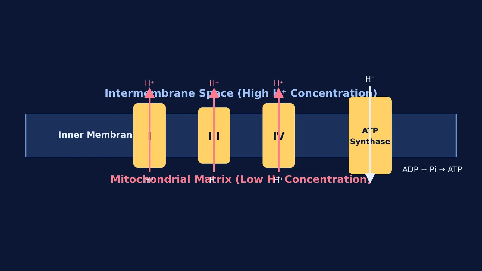
The electron transport chain: Electrons from donors like NADH and FADH₂ are passed through a series of protein complexes (Complexes I to IV) in the inner mitochondrial membrane. If you need a deeper look at how these proteins function, read our guide on the electron transport chain's Complexes I to IV.
Chemiosmosis: As electrons move through the chain, the complexes pump protons (H⁺) from the mitochondrial matrix into the intermembrane space. This creates a steep electrochemical gradient. The enzyme ATP synthase allows the protons to flow back into the matrix, using the kinetic energy to add a phosphate group to ADP, creating ATP.
How Lehninger changed bioenergetics
By isolating the function of mitochondria, Lehninger and Kennedy transformed biochemistry. Instead of studying whole cells or crushed tissue soup, researchers could now study energy production in pure, intact organelles. Lehninger went on to write *Principles of Biochemistry*, a textbook that has trained generations of scientists (Wikipedia).
If you are revising cell biology, you can ask Fermi's AI tutor to check your understanding of the enzymes of the citric acid cycle or the exact location of each process.
How to map cellular respiration for exams
Questions on the site of oxidative phosphorylation and respiration are standard in board exams and medical entrances. Unlike the chloroplast, which is the site of photosynthesis (see How to sketch and label a chloroplast for NEET biology), the mitochondrion handles respiration. Use this checklist to map where each stage happens (Wikipedia):
Glycolysis: Occurs in the cytoplasm. It requires no oxygen and produces a net of two ATP and two NADH molecules.
Link Reaction: Occurs in the mitochondrial matrix. Pyruvate is converted to Acetyl-CoA.
Krebs Cycle (TCA Cycle): Occurs in the mitochondrial matrix. It produces NADH, FADH₂, ATP (or GTP), and carbon dioxide.
Oxidative Phosphorylation: Occurs on the inner mitochondrial membrane. This is where the NADH and FADH₂ are cashed in to produce the bulk of the cell's ATP.
By memorising the location of each step, you can easily avoid the common traps in multiple-choice questions that mix up the matrix, the inner membrane, and the intermembrane space.