

On this page
Why is the chloroplast diagram tested so heavily in NEET?
In competitive examinations like NEET and CBSE board papers, the chloroplast is rarely tested as an isolated drawing. Instead, examiners use this organelle to verify whether you understand how cellular architecture drives bioenergetics. Under Class 11 biology specifications outlined by the National Council of Educational Research and Training, typical photosynthetic plant chloroplasts measure 5 to 10 µm in length and 2 to 4 µm in width. Mesophyll cells typically house 20 to 40 chloroplasts each, whereas the unicellular alga *Chlamydomonas* contains just 1 per cell.
Historically, the recognition that chloroplasts contain distinct internal compartments began in 1883, when German botanist Arthur Meyer published *Das Chlorophyllkorn in chemischer, morphologischer und biologischer Beziehung*. Meyer observed that chlorophyll is not uniformly distributed throughout the organelle, but concentrated in discrete green granules he named "grana", embedded within a colourless matrix known as the stroma. To explore how organelle specialization fits into cellular classification, see our review of Cell: the unit of life for NEET.
How should you draw the envelope and internal membrane system?
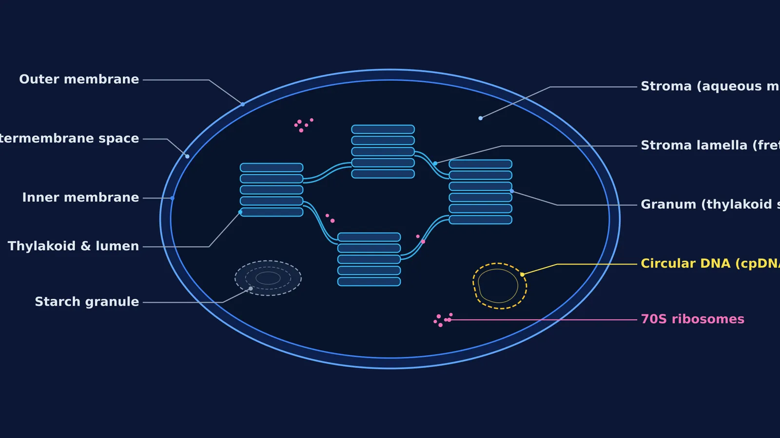
When sketching a chloroplast in an examination, start with its double-membrane envelope before adding internal details:
Outer membrane: A smooth lipid bilayer containing channel-forming porins that make it freely permeable to small molecules and ions.
Inner membrane: A selectively permeable barrier regulating metabolite transport into the stroma.
Intermembrane space: The narrow aqueous gap between the outer and inner boundaries.
Inside the inner membrane lies the thylakoid network. Wilhelm Menke coined the term "thylakoid" in 1962 in the Annual Review of Plant Physiology from the Greek *thylakos*, meaning sac or pouch. Thylakoids are flattened, disc-shaped membranous sacs stacked like coins into grana (singular: granum). High-resolution cryo-electron tomography by Benjamin D. Engel and colleagues in 2015 demonstrated that the thylakoid lumen measures roughly 9 nm across, forming a continuous internal aqueous space throughout the entire membrane system.
Connecting distinct grana across the stroma are flat, unstacked membranous tubules called stroma lamellae (or intergranal frets).
What is the difference between grana and stroma lamellae?
NEET frequently tests the biochemical division of labour between stacked grana and unstacked stroma lamellae:
Grana thylakoids contain both Photosystem I (PS I) and Photosystem II (PS II). They drive non-cyclic photophosphorylation, splitting water on the luminal face to release oxygen and generating both ATP and NADPH.
Stroma lamellae lack Photosystem II and NADP reductase. Operating with PS I alone, they perform cyclic photophosphorylation, generating ATP without producing NADPH or oxygen.
This spatial separation prevents excitation energy from spilling uncontrollably between complexes. Peter Mitchell's chemiosmotic hypothesis, published in Nature in 1961, showed that protons pumped into the thylakoid lumen generate an electrochemical gradient driving CF₀-CF₁ ATP synthase facing the stroma.
In the surrounding stroma, the enzyme RuBisCO fixes carbon dioxide into carbohydrates during the Calvin cycle. To see how enzyme kinetics influence carbon fixation, read our guides on why RuBisCO wastes energy in photorespiration and Blackman's law of limiting factors.
When reviewing cell biology concepts across botany, the Fermi Concept Library maps every concept in biology, bioenergetics, and cell structure, each with its own lesson and its place in the map.
Why must your sketch include 70S ribosomes and circular DNA?
A complete chloroplast sketch must include its non-membranous stroma inclusions:
Chloroplast DNA (cpDNA): A naked, circular, double-stranded DNA molecule lacking histone proteins, resembling bacterial plasmids.
70S ribosomes: Prokaryotic-type ribosomes consisting of 50S and 30S subunits, distinctly smaller than cytoplasmic 80S ribosomes.
Starch granules and plastoglobules: Stored photosynthetic carbohydrates and lipid droplets suspended in the stroma.
These inclusions prove that chloroplasts are semi-autonomous organelles capable of transcribing RNA and synthesizing several of their own core photosynthetic proteins. This structural evidence supports the endosymbiotic theory, showing chloroplasts evolved from ancient cyanobacteria engulfed by ancestral eukaryotes.
If you find yourself memorizing labels without grasping the underlying mechanisms, Fermi's AI tutor can work out what you already know and build a plan for the session, holding a line of reasoning and checking understanding as you go.
What four-step routine ensures full marks in board exams?
Follow this four-step routine to produce a clean, accurate sketch under timed exam conditions:
Establish the envelope: Draw an elongated ellipse across your answer sheet, then add a parallel inner contour just inside it to mark the outer membrane, inner membrane, and intermembrane space.
Assemble grana stacks: Within the stroma cavity, sketch three to four vertical stacks of flattened thylakoid discs aligned like coins.
Bridge with stroma lamellae: Connect adjacent stacks with thin, double-walled horizontal tubules spanning between discs.
Populate and label: Add a looped strand for circular DNA, fine stippling for 70S ribosomes, and an oval starch grain. Rule straight, horizontal leader lines to the margin and write labels in neat block letters.


