On this page
Human development unfolds through a strictly timed sequence of cell divisions, tissue foldings, and anatomical maturation. For students preparing for Class 12 biology boards and NEET-UG, few topics cause as much recurring confusion as the distinctions between a zygote, an embryo, and a foetus. Developmental embryology assigns each term to an exact biological stage marked by definite chronological boundaries and cellular milestones.
What happens during the zygote stage?
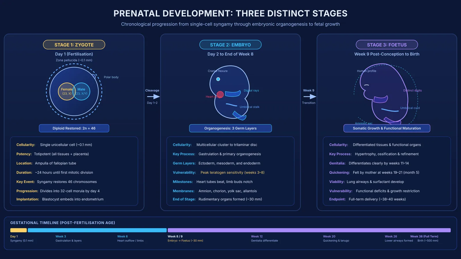
A zygote is the single diploid cell formed when a haploid sperm (23 chromosomes) fuses with a haploid secondary oocyte (23 chromosomes). This process, known as syngamy, occurs within 24 hours of ovulation in the ampulla of the four parts of the fallopian tube. According to a StatPearls review of fertilisation and cleavage by Rebecca Oliver and Hajira Basit, syngamy restores the complete human chromosome complement of 46 chromosomes (2n = 46), creating a unique genetic blueprint.
As a totipotent cell, the zygote can generate all embryonic tissues and the placenta. Within 24 hours of fertilisation, it initiates cleavage—rapid mitotic divisions partitioning cytoplasm into blastomeres without enlarging the conceptus. You can review how chromosome numbers remain constant during these divisions in our guide to chromosome counts through mitosis. As Oliver and Basit document in their StatPearls study on early cell division, cleavage produces a compact 32-cell morula by day 4. By day 5, fluid accumulates inside to form a blastocyst, which subsequently implants into the maternal endometrium.
How does an embryo develop during organogenesis?
The term embryo refers to the developing organism from first cleavage through the end of week 8 post-fertilisation. Once the blastocyst implants, its inner cell mass reorganises. During week 3, gastrulation converts the bilaminar disc into a trilaminar embryo. As detailed in the StatPearls guide to gastrulation by Jeremy Muhr, Tafline C. Arbor, and Kristin M. Ackerman, gastrulation establishes the three primary germ layers—ectoderm, mesoderm, and endoderm—as well as the primary body axes:
Ectoderm forms the nervous system and epidermis.
Mesoderm forms the cardiovascular system, skeleton, muscles, kidneys, and gonads.
Endoderm develops into the lining of the digestive and respiratory tracts.
Simultaneously, extraembryonic membranes protect and nourish the embryo, as explored in our guide to the functions of the amnion, chorion, yolk sac and allantois. During weeks 6 through 8, the cardiovascular system remodels—including outflow tract division into the aorta and pulmonary artery—while limbs lengthen and facial contours emerge. According to developmental records catalogued in StatPearls by Mary F. Donovan and Marco Cascella, this rapid differentiation makes weeks 3 through 8 the most vulnerable window for teratogenic disruption.
When does an embryo become a foetus?
The transition from embryo to foetus occurs at the start of week 9 post-fertilisation (the start of month 3). As documented in a StatPearls analysis of embryonic weeks 6 through 8 by Mary F. Donovan and Marco Cascella, organogenesis is fundamentally complete by the end of week 8. At this transition, the organism displays a distinctly human profile and possesses the rudimentary layout of every major organ system.
From week 9 until birth (around week 38 post-fertilisation, or week 40 of clinical gestation), the developing organism is classified as a foetus. Growth and tissue refinement replace initial organ formation. According to the MedlinePlus medical encyclopedia overview of fetal development, milestones follow a reliable schedule:
By weeks 11 to 14 of gestation (around week 12), facial features form, external genitals appear, and the baby can make a fist.
During weeks 19 to 21 (month 5), quickening occurs, lanugo appears, and swallowing begins.
Between weeks 23 and 26, lung airways develop and bone marrow produces blood cells, preceding third-trimester surfactant synthesis.
How can you tell the three stages apart in exams?
When tackling exam questions, use this 4-point framework to distinguish each stage:
Cellular organisation: A zygote is unicellular. An embryo is a multicellular cluster or folded disc forming germ layers and organ primordia. A foetus is an anatomically recognisable organism with established organ systems.
Primary developmental mechanism: Cleavage and blastulation define the zygote; organogenesis defines the embryo; somatic growth and tissue maturation define the foetus.
Chronological timeline: As clinical texts referenced by the National Library of Medicine confirm, the zygote spans Day 1; the embryo spans Day 2 through Week 8 post-fertilisation; the foetus spans Week 9 post-fertilisation through birth.
Pathological vulnerability: Pre-implantation exposures cause an all-or-none response. Embryonic exposures (weeks 3 to 8) cause major structural malformations, whereas fetal exposures primarily produce functional deficits or growth restriction.
Visualising the transformation from a single cell into layered tissues can feel abstract from static textbook figures. Students seeking to master embryogenesis can consult the Fermi Concept Library, which maps every concept in biology and clarifies cell division pathways alongside organ development. When working through complex assertion-reason questions or multi-step genetics problems, the step-by-step solver in the Fermi Problem Library explains the underlying biological reasoning at each stage rather than simply printing a solution.