

On this page
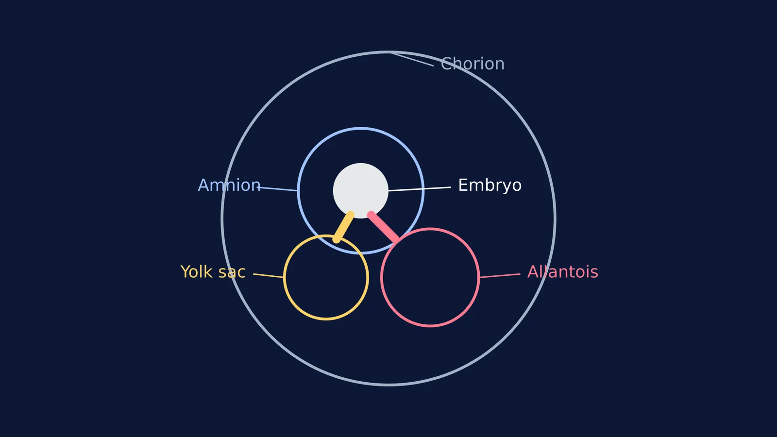
When an embryo develops, it does not grow alone. In mammals, birds and reptiles, it is supported by four extra-embryonic membranes: the amnion, chorion, yolk sac and allantois. Together, these tissues protect the growing embryo and manage its life-support systems, from exchanging gases to transferring nutrients.
The development of these membranes marked a major evolutionary leap. Fish and amphibians are anamniotes; they lay their eggs in water and do not develop these protective layers. The evolution of the amnion and its accompanying membranes allowed early vertebrates to leave the water and reproduce on dry land, defining the entire clade known as amniotes. Understanding how they work is a cornerstone of embryology. If you want to master these concepts, the Fermi Concept Library explains every concept in biology with its own lesson and its place in the map.
What are the functions of the amnion and chorion?
The amnion is the inner membrane that closely covers the early embryo, developing from the extra-embryonic somatic mesoderm and ectoderm. As it develops, it fills with amniotic fluid, expanding into what is commonly known as the amniotic sac. This fluid acts as a crucial physical cushion, providing a protective, buoyant environment where the fetus can move freely within the walls of the uterus. During labour, the spontaneous rupture of this sac is what is referred to as "waters breaking". Medical professionals also sample this fluid between 15 and 20 weeks of pregnancy during a procedure called amniocentesis to check for chromosomal abnormalities.
The chorion is the outermost fetal membrane. In humans and other mammals, its primary job is to bridge the gap between the mother and the developing fetus. The chorion completely surrounds the amnion and the embryo, acting as a protective barrier and the main interface for nutrient exchange.
How do chorionic villi form the placenta?
As the chorion grows, it rapidly proliferates to form finger-like projections called chorionic villi. These villi are initially small and non-vascular, consisting only of trophoblast cells. However, they soon increase in size and ramify as they invade and destroy the uterine decidua (the lining of the uterus) to absorb nutritive materials.
As they branch out, the villi become vascularised by branches of the umbilical vessels, specifically the umbilical arteries and vein. This allows the chorion to transfer nutrients and oxygen from the maternal blood into the fetal blood, and move waste products in the opposite direction. Initially, villi cover the entire chorion, but eventually, those in contact with the decidua capsularis atrophy to become the smooth *chorion laeve*. Meanwhile, the villi at the embryonic pole increase greatly in size and complexity to form the *chorion frondosum*, which fuses with the maternal tissue (decidua basalis) to create the mature placenta. To trace how the embryo reaches this critical stage in the uterus, you can review From fertilisation to implantation: the first week of pregnancy.
What do the yolk sac and allantois do?
In birds and reptiles, the yolk sac holds the nutrient-rich yolk that feeds the embryo throughout its development inside the shell. However, in humans and most mammals, the embryo gets its long-term nutrition from the placenta. Despite this shift in function, the yolk sac remains crucial in early development. It is formed by cells of the hypoblast layer and is responsible for the early embryonic blood supply before organs like the liver and bone marrow take over. Much of the human yolk sac is eventually incorporated into the primordial gut during the fourth week of embryonic development.
The allantois is a hollow, sac-like structure filled with clear fluid. It extends from the early yolk sac and helps the embryo handle liquid waste and exchange gases. In reptiles and birds, it fuses with the chorion to form the chorioallantoic membrane, which breathes through the porous eggshell. In humans, as the embryo grows, the blood vessels of the allantois become the umbilical arteries and vein, physically connecting the fetus to the placenta and ensuring a constant flow of resources.
The complex interplay of these membranes—how they fold, fuse and direct the fate of cells—was a central puzzle for early developmental biologists. Some of the most famous experiments in this field were conducted by researchers studying how embryonic cells communicate, such as those by Hans Spemann, Hilde Mangold and the embryo organizer.
A framework for remembering membrane functions
When revising these four structures, it helps to match each membrane to its everyday biological equivalent:
Amnion = The shock absorber. It creates the fluid-filled sac that cushions and protects, providing buoyancy and room for movement.
Chorion = The trading post. It forms the outer boundary and the chorionic villi, merging with maternal tissue to build the placenta for exchange.
Yolk sac = The early blood factory. It kickstarts blood cell production before the embryo's organs have fully formed.
Allantois = The waste pipe and cable. It manages early liquid waste, and its vessels ultimately form the umbilical cord.


