

On this page
Every month, an ovary releases an egg. But the ovary is not directly connected to the uterus. Instead, the egg must be caught and transported by the fallopian tube (also known as the oviduct). The fallopian tube is a muscular channel that is 10 to 14 cm long. This tube is where the sperm and egg finally meet.
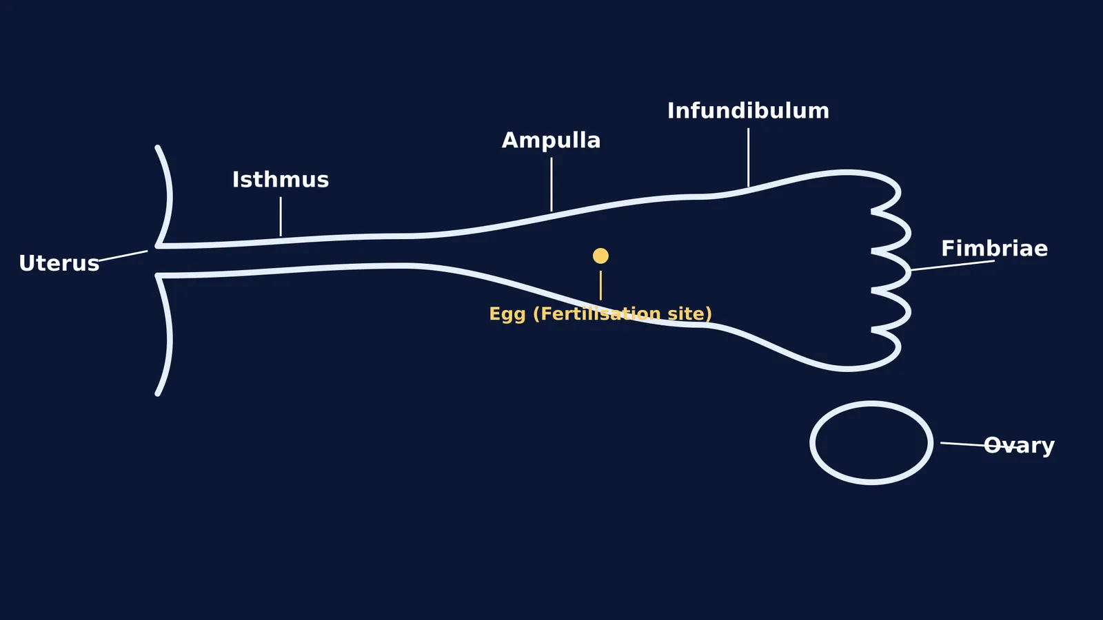
For medical entrance exams like NEET, you need to know the anatomy of this tube in order. From the ovary to the uterus, the fallopian tube is divided into four distinct regions: the fimbriae, the infundibulum, the ampulla and the isthmus.
How do the fimbriae and infundibulum catch the egg?
When ovulation occurs, the egg is released into the open space of the abdominal cavity. The infundibulum is the funnel-shaped distal opening of the fallopian tube that sits just above the ovary.
To make sure the egg does not get lost, the edges of the infundibulum feature fringe-like projections called fimbriae. These projections are typically about 1 mm wide. They sweep over the ovary like small tentacles. During ovulation, the fimbriae move closer to the ovary and create fluid currents that draw the released egg safely into the infundibulum. One specific, longer projection called the ovarian fimbria maintains constant contact with the ovary to guide the process.
Where does fertilisation usually take place?
Once inside the tube, the egg moves into the ampulla. The ampulla is the longest section of the tube, measuring about 5 cm in length and up to 1 cm in width.
The ampulla is the most critical site for human reproduction because fertilisation usually takes place in the ampulla. If sperm are present in the female reproductive tract, they swim up through the uterus and into the fallopian tube to meet the egg here.
The walls of the ampulla are highly folded and lined with two key types of cells:
Ciliated cells: These have tiny hair-like structures that beat in waves, sweeping the egg (or the newly formed zygote) towards the uterus.
Peg cells: These are non-ciliated secretory cells that release a fluid rich in nutrients, providing a safe environment to sustain both the egg and the sperm.
The isthmus: the final transport zone
After the ampulla, the tube narrows significantly into the isthmus. The isthmus is a thick-walled, cord-like section that is approximately 3 cm long.
Because it is much narrower (only 1 to 5 mm wide) and has a stronger muscular layer, the isthmus acts as a transport channel and a temporary reservoir for sperm. Once fertilisation happens in the ampulla, it takes the fertilised egg several days to travel through the isthmus and the short intramural part (the 1 cm segment inside the uterine wall) before it finally drops into the uterine cavity for implantation.
The muscles that drive the journey
While the cilia brush the egg along, the tube itself physically pushes it. The middle layer of the fallopian tube, known as the muscularis, consists of an inner circular and outer longitudinal layer of smooth muscle. These muscles generate rhythmic, wave-like contractions called peristalsis that propel the egg towards the uterus.
This is also the part of the anatomy involved in tubal ligation. This surgical method of permanent birth control involves blocking or cutting the fallopian tubes to prevent the sperm from ever reaching the ampulla.
How to remember the sequence for exams
If you struggle to remember the order of these parts for biology tests, follow the path of the egg from outside to inside:
Fimbriae: Catch the egg.
Infundibulum: The funnel entry.
Ampulla: The wide middle where sperm meets egg.
Isthmus: The narrow bridge to the uterus.
If you are revising reproductive anatomy and find it hard to picture how these structures link together, the Fermi Concept Library can help. The AI tutor can draw out the female reproductive system and quiz you on the functions of each part, checking your understanding as it goes.
For more on what happens after the egg reaches the uterus, you can read about From fertilisation to implantation: the first week of pregnancy, or explore the final stages of the reproductive cycle in Parturition and lactation: the hormones behind birth and milk.


