

On this page
Whiskers are far more than ordinary fur; they are specialized tactile antennas that map a cat’s immediate environment. Known scientifically as vibrissae—from the Latin *vibrare*, meaning to vibrate—these stiff hairs allow felines to navigate darkness, evaluate tight spaces, and track prey with rapid precision.
How do cat whiskers work as sense organs?
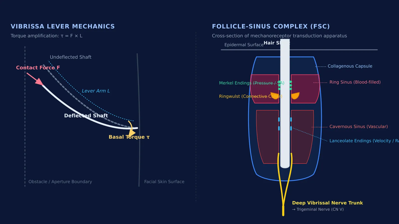
The whisker shaft itself is composed of dead, keratinized cells without any nerve endings. All sensory detection occurs at the base, where each hair is anchored within a specialized follicle-sinus complex (FSC) embedded deep into the skin.
Inside this structure, the follicle is enveloped by a collagenous capsule containing two blood-filled spaces: an upper ring sinus and a lower cavernous sinus. In *The Journal of Comparative Neurology*, Rice, Mance, and Munger (1986) identified six distinct mechanoreceptor populations within this feline complex, including slowly adapting Merkel cell-neurite complexes at the ring sinus and longitudinally aligned lanceolate endings.
When external contact or an air current deflects the whisker, the shaft acts as a mechanical lever. The movement displaces blood within the sinuses, concentrating hydraulic pressure onto the ringwulst—a dense connective tissue collar that focuses mechanical strain onto the sensory nerve endings. Confocal analyses by Ebara et al. (2002) confirmed that these dense receptor clusters transmit rich tactile signals directly into the trigeminal nerve system.
Why do cats have whiskers on their cheeks, eyes, and legs?
A cat’s vibrissae are organized into distinct anatomical groups across the body:
Mystacial vibrissae: Prominent whiskers arranged in horizontal rows across the muzzle, used for navigation and measuring gaps.
Superciliary vibrissae: Positioned above the eyes to trigger an automatic protective blink reflex.
Genal and mandibular vibrissae: Situated on the cheeks and chin to monitor lateral and ground clearance.
Carpal vibrissae: Located on the underside of the front wrists (carpus).
Carpal vibrissae resolve a major limitation in feline vision. While domestic cats excel in low light, their large, fixed-focus lenses struggle to accommodate objects positioned directly beneath the mouth and paws, creating an immediate visual blind spot. In *The Journal of Physiology*, Gottschaldt, Iggo, and Young (1973) recorded afferent signals from carpal sinus hairs through the ulnar nerve, demonstrating that slowly adapting units and rapidly adapting Pacinian corpuscles track struggling prey to guide a precise bite without visual input.
How do whiskers detect air currents and narrow gaps?
The sensitivity of feline vibrissae stems from lever mechanics. When a lateral contact force F acts at distance L along the shaft, it generates a basal bending torque τ = F × L. Because the shaft is stiff and tapers toward the tip, even a microscopic deflection produces significant torque and fluid displacement within the blood sinus.
In *The Journal of Neurophysiology*, Dykes (1975) found that 66% of afferent fibers in the cat’s infraorbital nerve serve vibrissae. Two-thirds are rapidly adapting fibers that detect velocity and vibration, while slowly adapting fibers convey 2.7 bits of spatial data for deflections under 17 degrees.
This sensory machinery powers two key behaviours:
Air current detection: As a cat moves forward, its body displaces air that bounces off walls and solid objects. The whiskers detect these reflected micro-drafts, functioning like tactile sonar in the dark.
Gap sizing: Mystacial whiskers spread to roughly the width of the cat’s body. If the whiskers pass through an opening without bending, the cat knows its shoulders and torso will fit.
What happens during whisker stress and bowl fatigue?
"Whisker fatigue" describes the popular theory that deep, narrow food dishes cause sensory stress through repetitive contact with the whiskers.
To evaluate this, Slovak and Foster (2021) tested 40 indoor cats in the *Journal of Feline Medicine and Surgery*. Comparing deep dishes with shallow, whisker-friendly alternatives, they observed no significant change in eating time (P = 0.8) or amount eaten (P = 0.7). However, 74% of cats preferred the shallow dishes when given a choice. While not a clinical illness, wide bowls minimize unnecessary mechanical stimulation of facial nerves.
How do mechanoreceptors connect to neural coordination?
For students examining neurobiology, feline vibrissae demonstrate core sensory principles:
Receptor types: Merkel discs provide slowly adapting responses to sustained pressure, whereas Pacinian corpuscles offer rapidly adapting responses to vibrations.
Sensory transduction: Mechanical displacement deforms the receptor membrane, opening mechanosensitive ion channels. The resulting sodium influx generates a receptor potential that triggers action potentials along myelinated axons. You can explore how impulses travel in our guide to nerve impulse propagation and reflex arcs.
Neural mapping: Trigeminal afferents project to dedicated cortical barrels in the primary somatosensory cortex, demonstrating the somatotopic maps explored in our profile of Wilder Penfield and cortical brain maps.
Visualizing biophysical forces and ionic gradients is often challenging from static diagrams alone. The Fermi Simulation Lab offers interactive simulations for the ideas that are easier to see than to read. You can also explore the Fermi Concept Library for every concept in physics, chemistry, biology, mathematics and computing, each with its own lesson and its place in the map, or review reflex pathways in our primer on Class 10 control and coordination.


