

On this page
When you catch a falling glass, your heart races and your fingers snap shut. Although these responses seem simultaneous, your nervous system processes them through different pathways. Human actions divide into three categories: voluntary actions guided by conscious choice, involuntary actions sustaining life in the background, and reflex actions executing rapid protective manoeuvres without prior deliberation. Telling them apart is essential in human biology, notably within the Class 10 control and coordination syllabus outlined by the National Council of Educational Research and Training.
Everyday speech often calls any automatic motion a reflex. In reality, all reflex actions are involuntary, but not all involuntary actions are reflexes. Involuntary actions, such as cardiac contraction or intestinal peristalsis, run continuously through autonomic circuits to maintain homeostasis. In contrast, reflex actions are discrete motor responses triggered by acute sensory stimuli. Voluntary actions, such as writing notes or kicking a ball, stem from conscious intention.
Which parts of the nervous system control each action?
Each category of action relies on a dedicated anatomical control centre and specialised conduction pathways:
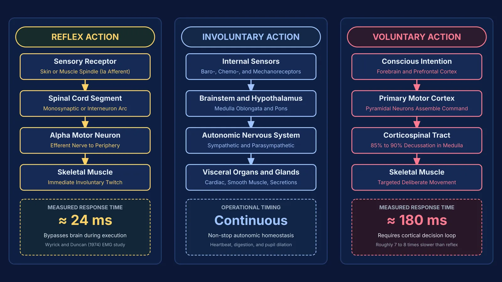
Voluntary actions originate in the cerebral cortex of the forebrain, mainly within the primary motor cortex. As detailed by AbuHasan and Munakomi (2023) in StatPearls, voluntary signals descend through the pyramidal tract, where roughly 85% to 90% of corticospinal fibres decussate in the medulla oblongata before synapsing on lower motor neurons that innervate skeletal muscles.
Involuntary actions are coordinated subcortically by the brainstem, especially the medulla oblongata and pons, under hypothalamic modulation. According to StatPearls research on autonomic physiology by LeBouef, Yaker, and Whited (2023), the autonomic nervous system uses sympathetic and parasympathetic divisions to regulate smooth muscle, cardiac tissue, and glands.
Reflex actions bypass conscious brain circuits during execution. As documented by Zimmerman and Hubbard (2023), a classical stretch reflex forms a localised reflex arc where sensory afferent neurons enter the spinal cord's dorsal horn and synapse directly onto alpha motor neurons in the anterior horn, contracting muscle before ascending signals reach the cortex.
When mapping how distinct brain regions coordinate complex behaviours, the Fermi Concept Library covers every concept in biology, each with its own lesson and its place in the map. To explore how the forebrain and hindbrain divide these responsibilities, review our overview of the roles of the forebrain, midbrain, and hindbrain.
Why are reflex actions faster than voluntary actions?
The evolutionary purpose of a reflex is survival speed. If your foot steps on a nail, waiting for signals to ascend the spinal cord, reach the cortex, generate a plan, and descend back would permit deep tissue damage. By routing the motor command directly through the spinal cord, the reflex arc eliminates precious milliseconds of synaptic delay.
This speed difference is measurable through electromyography. In an experimental study involving 30 participants subjected to sudden joint displacement, Wyrick and Duncan (1974) observed an average spinal reflex latency of 24 milliseconds, while voluntary simple reaction times averaged 180 milliseconds. That physiological shortcut makes the spinal reflex roughly 7 to 8 times faster than a deliberate motor response. Furthermore, neurophysiological research by Forgaard et al. (2015) in the Journal of Neurophysiology demonstrated that short-latency reflex responses occur within 50 milliseconds, whereas voluntary motor outputs typically require over 100 milliseconds to assemble cortical motor programs. For a deeper look at action potentials and synaptic transmission times, read our guide on nerve impulse transmission and reflex arcs.
How can you classify an action in three diagnostic steps?
When analyzing physiological scenarios on exams or in everyday life, you can pinpoint any action using a three-step decision framework:
Test for conscious volition: Can you choose when to initiate, pause, or alter the speed of the action? If yes, the action is voluntary and governed by the cerebral cortex acting on somatic muscles.
Test for an acute sensory trigger: Did the movement occur as an instantaneous, stereotypic jerk provoked by an abrupt external stimulus? If yes, it is a reflex action executed through a spinal or cranial reflex arc.
Test the target organ and continuity: Does the action operate continuously on cardiac tissue, smooth muscle, or glandular secretions without conscious awareness? If yes, it is an involuntary autonomic function driven by the brainstem.
Boundary cases demonstrate how these circuits interact. Swallowing begins voluntarily as the tongue propels food backward, but once the bolus touches the pharyngeal wall, the medulla takes over involuntary peristalsis. Similarly, blinking can be voluntary when winking, involuntary when hydrating the cornea, or a protective reflex when dust nears the eye. When tackling multi-part problems on neural mechanisms, the Fermi Problem Library offers worked problems with a step-by-step solver that explains the reasoning rather than printing an answer.


