On this page
When an electrical signal reaches the end of a nerve cell, it cannot simply jump the gap to the next cell. Instead, the signal is converted into a chemical message. The junction where this happens is called a chemical synapse.
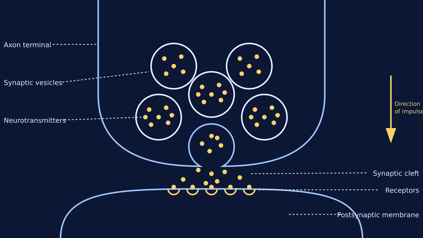
Whether you are studying for NEET biology or your Class 11 exams, the synapse is a high-yield topic. A standard synapse diagram breaks this microscopic junction into three main zones: the sending side, the gap, and the receiving side.
What is the structure of a synapse diagram?
The structure of a chemical synapse is asymmetric, meaning the two sides look completely different. A complete diagram includes these distinct parts:
Axon terminal (Presynaptic membrane): The bulb-like ending of the sending neuron. It is packed with mitochondria to provide energy and contains the machinery needed to release chemical signals.
Synaptic cleft: The physical gap between the two cells. The synaptic cleft is approximately 20 nanometres (nm) wide. This tiny volume ensures that a small amount of chemical can cause the concentration to spike and drop rapidly, enabling incredibly fast communication between neurons.
Postsynaptic membrane: The surface of the receiving cell, which is usually a dendrite or a muscle fibre. It is thickly packed with receptor proteins that are specifically shaped to bind with the incoming signal.
If you want to see how this junction fits into the wider nervous system, you can explore the Concept Library to map out the connections between the brain and the rest of the body.
How do synaptic vesicles release neurotransmitters?
The chemical messengers themselves are called neurotransmitters. To protect them from being broken down by enzymes in the cytoplasm, the axon terminal stores them in small bubbles called synaptic vesicles. These vesicles are tiny, membrane-bound spheres that are approximately 40 nm in diameter.
The release process, called exocytosis, follows a strict, calcium-dependent sequence:
An electrical impulse, known as an action potential, arrives at the axon terminal.
This sudden voltage change opens voltage-gated calcium channels, allowing calcium ions to flood into the terminal from the surrounding fluid.
The calcium acts as a crucial trigger, causing the synaptic vesicles to move to the active zones at the edge of the presynaptic membrane.
The vesicle membrane fuses with the cell membrane, spilling its neurotransmitters into the synaptic cleft.
Once in the cleft, the neurotransmitters drift across and lock into specific receptors on the postsynaptic membrane, which can either trigger or inhibit a new electrical signal. You can read more about how these chemicals differ from endocrine signals in our guide to Neurotransmitter vs Hormone.
Why is the direction of transmission one-way?
A frequent exam question asks why nerve impulses only travel in one direction across a synapse. The answer lies directly in the diagram's asymmetry.
The machinery required to send a signal—specifically the neurotransmitter-filled synaptic vesicles and the voltage-gated calcium channels—only exists in the presynaptic axon terminal. Conversely, the machinery needed to receive the signal—the specialized protein receptors—only exists on the postsynaptic membrane. Because neurotransmitters can only be released from one side and can only bind to the other, the signal absolutely cannot go backwards. This one-way valve effect ensures that signals travel in orderly, predictable circuits, such as the pathways connecting Sensory vs motor neurons.
How to draw a synapse diagram for exams
Drawing a chemical synapse from memory is a standard biology exam task. Use this sequence to get the proportions and labels right without wasting time during your test:
Draw the bulb: Start with a large, inverted U-shape to represent the axon terminal. Make it wide enough to fit several circles inside.
Draw the receiving membrane: Below the bulb, draw a parallel curve. Leave a clear, even gap between them to represent the synaptic cleft.
Add the vesicles: Inside the bulb, draw five or six small circles.
Show exocytosis: Draw an omega shape (Ω) on the bottom edge of the bulb to show one vesicle fusing with the presynaptic membrane and opening into the cleft.
Add the neurotransmitters: Draw small dots inside the unbroken vesicles, spilling out of the fused vesicle, floating in the cleft, and sitting on the receiving membrane.
Add the receptors: Draw small cup-like shapes on the postsynaptic membrane exactly where the neurotransmitter dots are sitting.
If you are ever unsure about the structural differences between nerve cells, such as Multipolar, bipolar and unipolar neurons, sketching their synaptic connections can make their distinct functions much easier to visualise and remember.