

On this page
Every neuron in the human nervous system shares the same fundamental task of transmitting electrical signals, yet their physical architectures diverge into distinct classes. In foundational biology syllabi and medical histology, structural classification depends on counting the protoplasmic extensions, or neurites, that emerge directly from the soma (cell body). According to the NCERT Class 11 Biology curriculum, neurons fall into three primary morphological groups: unipolar, bipolar, and multipolar. A comprehensive anatomical review in StatPearls reinforces that this structural variation directly shapes how individual cells collect, integrate, and dispatch neural information throughout the central and peripheral nervous systems.
Understanding this framework is essential not only for scoring full marks in competitive entrance tests like NEET-UG, but also for appreciating how cellular anatomy underpins complex neurobiology. For a broader overview of impulse transmission across synapses and reflex circuits, read our neural control and coordination guide.
How do you classify neurons by their number of processes?
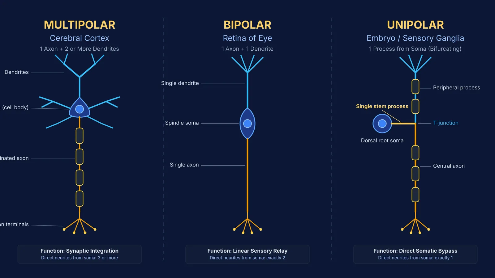
The structural classification of neurons evaluates the polarity of the cell body by tallying its direct cytoplasmic projections:
Unipolar neurons: A single process originates from the cell body and functions as an axon without separate dendrites. In human physiology, true unipolar neurons are predominantly restricted to early embryonic development.
Bipolar neurons: Two distinct processes emerge from opposite poles of the soma—one dedicated dendrite that receives input and one axon that carries output away.
Multipolar neurons: One single axon accompanies two or more branching dendrites projecting from various regions of the cell body membrane.
Why does the cerebral cortex rely on multipolar neurons?
Multipolar neurons represent the overwhelming majority of nerve cells in the central nervous system. In the human brain, they populate the grey matter of the functional regions of the human brain, including the cerebral cortex, hippocampus, and cerebellar cortex, as well as the ventral horn of the spinal cord.
The structural hallmark of a multipolar cell is its elaborate dendritic tree. A seminal study by Spruston (2008) in *Nature Reviews Neuroscience* notes that pyramidal neurons make up roughly 70% to 80% of all cortical neurons. These cells feature multiple basal dendrites spreading locally and a prominent apical trunk ascending toward superficial cortical layers. This vast surface allows a single cortical neuron to receive and integrate synaptic inputs from tens of thousands of upstream partners through spatial and temporal summation before generating an action potential at the axon hillock. When navigating intricate neuroanatomy, the Fermi Concept Library covers every concept in biology, each with its own lesson and its place in the map.
Where are bipolar neurons found and how do they function?
Bipolar neurons are specialized for linear, high-fidelity sensory relay. Because they feature exactly one dendrite and one axon extending 180° apart from a spindle-shaped soma, they act as vertical conduits rather than computational integrators.
The classic textbook location for bipolar neurons is the retina of the eye. A landmark review by Euler et al. (2014) in *Nature Reviews Neuroscience* explains that retinal bipolar cells serve as the primary vertical bridge connecting light-sensing photoreceptors (rods and cones) in the outer plexiform layer to ganglion cells in the inner plexiform layer. Mouse and primate retinas contain at least 13 distinct functional types of bipolar cells that segregate visual signals into parallel ON and OFF channels without diluting spatial resolution. Bipolar neurons also occur in the olfactory epithelium and the spiral and vestibular ganglia of the vestibulocochlear nerve.
What happens to unipolar neurons after the embryonic stage?
During early neurogenesis, developing neuroblasts briefly present as unipolar cells with a solitary neurite. In sensory pathways, these cells differentiate into bipolar neuroblasts before undergoing a remarkable mechanical transformation.
As shown in historical neuroembryology and Cajal's histological studies of the neuron, the two opposing stems of sensory neuroblasts migrate toward one side of the soma and fuse together. This fusion creates a single T-shaped or Y-shaped stem process, yielding the pseudounipolar neuron typical of adult dorsal root ganglia and cranial nerve sensory ganglia. A computational modeling study by Sundt et al. (2015) in the *Journal of Neurophysiology* demonstrated that the morphological T-junction acts as an intrinsic low-pass filter, allowing peripheral touch and pain spikes to travel directly into the spinal cord while bypassing metabolic delay at the soma.
How can you solve neuron classification questions in exams?
NEET and board exam questions regularly test neuron types through matching columns or statement evaluations. To ensure you never drop marks on these questions, follow this 3-step decision checklist:
Count membrane origins: Look exclusively at the soma boundary. If 1 stem emerges, it is unipolar (embryo) or pseudounipolar (dorsal root ganglion); if 2 opposite stems emerge, it is bipolar (retina); if 3 or more stems emerge, it is multipolar (cortex).
Anchor the classic pairings: Memorize the NCERT tripartite standard: Multipolar = Cerebral Cortex; Bipolar = Retina of Eye; Unipolar = Embryonic Stage.
Verify the direction of flow: Remember that impulses travel through dendrites toward the soma (afferent) and through the single axon away from the soma (efferent).
For multi-statement matching questions and structural biology exercises, the Fermi Problem Library provides worked problems with a step-by-step solver that explains the reasoning rather than printing an answer.


