

On this page
If you accidentally touch a hot pan, you pull your hand away before you even realise you are in pain. That instant reaction relies on two distinct types of nerve cells working together seamlessly. The entire process happens in a fraction of a second, demonstrating the incredible efficiency of the human nervous system.
To understand the nervous system, you need to know exactly how a signal travels through the body. The distinction between sensory and motor neurons comes down to the direction of the electrical impulse, where their respective cell bodies sit, and what specific tissues they connect to each other.
What is the sensory neuron vs motor neuron difference?
The clearest difference is the direction in which they carry information through the peripheral nervous system. Sensory neurons act as the input system. They receive sensory information from the outside world or internal organs and transmit it to the central nervous system (OpenStax Biology). Because they bring signals towards the centre, they are called afferent neurons.
Motor neurons act as the output system. They transmit commands from the central nervous system outward to muscles, glands, or other target organs (OpenStax Biology). Because they carry signals away from the centre, they are called efferent neurons. To reach distant effectors, some motor neurons have axons up to one metre long in humans.
Where do their cell bodies sit?
The cell bodies, or somas, of these two types of neurons live in completely different anatomical locations relative to the spinal cord. This physical separation helps keep incoming and outgoing signals distinct.
The cell bodies of sensory neurons are located entirely outside the spinal cord. They are clustered together in a visible swelling called the dorsal root ganglion (Neuroscience, 2nd Ed.). Because these neurons are pseudounipolar, the electrical signal actually bypasses the cell body altogether, travelling directly from the peripheral axon in the limb to the central axon that enters the back of the spinal cord.
The cell bodies of motor neurons are located safely inside the central nervous system. Specifically, they sit in the ventral horn of the spinal cord's gray matter (Neuroscience, 2nd Ed.). From this protected location, their axons exit through the front of the spinal cord and travel all the way to the target muscle to trigger a contraction.
How do they form a reflex arc?
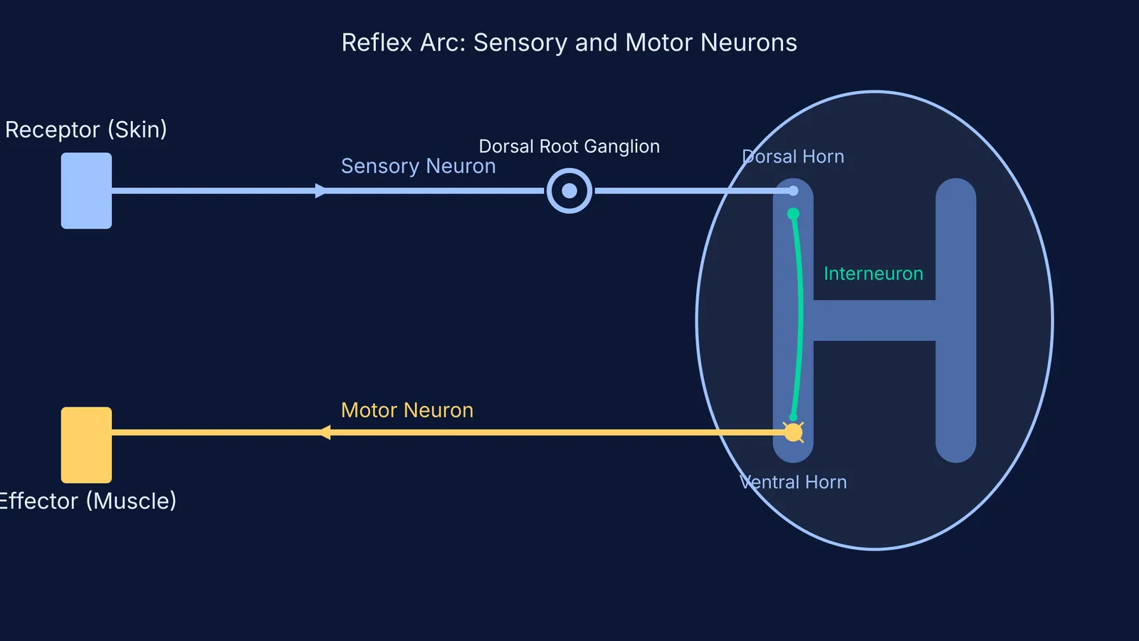
A reflex arc is the neural pathway that controls an involuntary reflex. By bypassing the conscious brain for immediate processing, the body can react to danger much faster than if the signal had to travel all the way up to the cerebral cortex and back down. It typically follows a specific, predictable path:
Receptor: A sensory receptor in the skin or tissue detects the environmental stimulus, such as heat or sharp pressure.
Sensory neuron: The nerve impulse travels up the long peripheral axon of the sensory neuron, passing its cell body in the dorsal root ganglion, and enters the dorsal horn of the spinal cord.
Interneuron: In most basic reflexes, the sensory neuron synapses directly onto a small relay cell called an interneuron located entirely within the spinal cord's gray matter.
Motor neuron: The interneuron immediately triggers a motor neuron sitting in the ventral horn.
Effector: The outgoing motor impulse travels down the axon to the arm or leg muscle, causing it to rapidly contract and pull your hand away from the danger.
If you find it hard to picture the anatomy from a textbook page, the Fermi Concept Library covers every concept in biology with clear, interactive diagrams. You can explore exactly how the signal flows through the reflex arc in real-time, while Fermi's AI tutor checks your understanding of the pathway as it goes.
A practical tool: The SAME DAVE mnemonic
When you are sitting an exam and suddenly doubt which root does what, it is easy to panic. To avoid confusion, use the classic medical mnemonic SAME DAVE:
Sensory = Afferent
Motor = Efferent
Dorsal = Afferent (Sensory signals always enter the dorsal root)
Ventral = Efferent (Motor signals always exit the ventral root)
Write this acronym down in the margin of your rough work as soon as the exam starts. It is a foolproof way to guarantee you will not mix up your input and output pathways when working under pressure.
If you are currently revising human physiology, comparing these control systems with other body networks helps lock in the core concepts. For example, look at how the formed elements of blood are distributed by the cardiovascular system, or review the divisions of the human skeleton to see exactly which specific bones those motor neurons ultimately move when they fire.


