On this page
The stomata slide in Class 10 looks simple, but small slips in it cost marks. CBSE's Class X Science curriculum for 2026-27 lists "Preparing a temporary mount of a leaf peel to show stomata" as Experiment 6, under Unit II, World of Living.
How do you prepare a temporary mount of a leaf peel?
You need a fresh leaf, a petri dish or watch glass of water, safranin, glycerine, a clean slide, a coverslip, a brush, a needle, blotting paper and a compound microscope. The NCERT Class X laboratory manual suggests a dicot leaf such as *Petunia*, *Dianthus* or *Solanum* (the genus that includes brinjal and tomato).
Fold or tear the leaf and pull off the thin, transparent skin from the lower surface. The NCERT manual calls it a "thin membranous transparent peel".
Drop the peel into water in a petri dish and add a drop of safranin.
Lift the stained peel onto the centre of a clean slide with a brush and add a drop of glycerine.
Lower the coverslip gently with a needle so no air bubbles get trapped.
Blot the extra liquid from the edges of the coverslip.
Focus under low power first, then switch to high power to see what is inside the guard cells.
Steps 1 to 4 are in the order that CBSE's Assessment of Practical Skills booklet for Class X marks correct in a sequencing question, and the 2026-27 curriculum lists that booklet as a prescribed book. The bubble, blotting and focusing advice comes from the NCERT manual, which mounts the peel in plain water; the stain and glycerine come from the CBSE booklet.
Why use safranin and glycerine?
Safranin is a stain that makes the cells easier to see. CBSE's answer key says "Safranin is pinkish red in colour", so expect a stained peel to look pinkish red under the microscope.
Glycerine stops the peel from drying out while you look at it. Educational guides to this practical give it the same job, and add two handling tips: move the peel with a brush so the cells do not tear, and blot away extra glycerine and stain.
What will you see under the microscope?
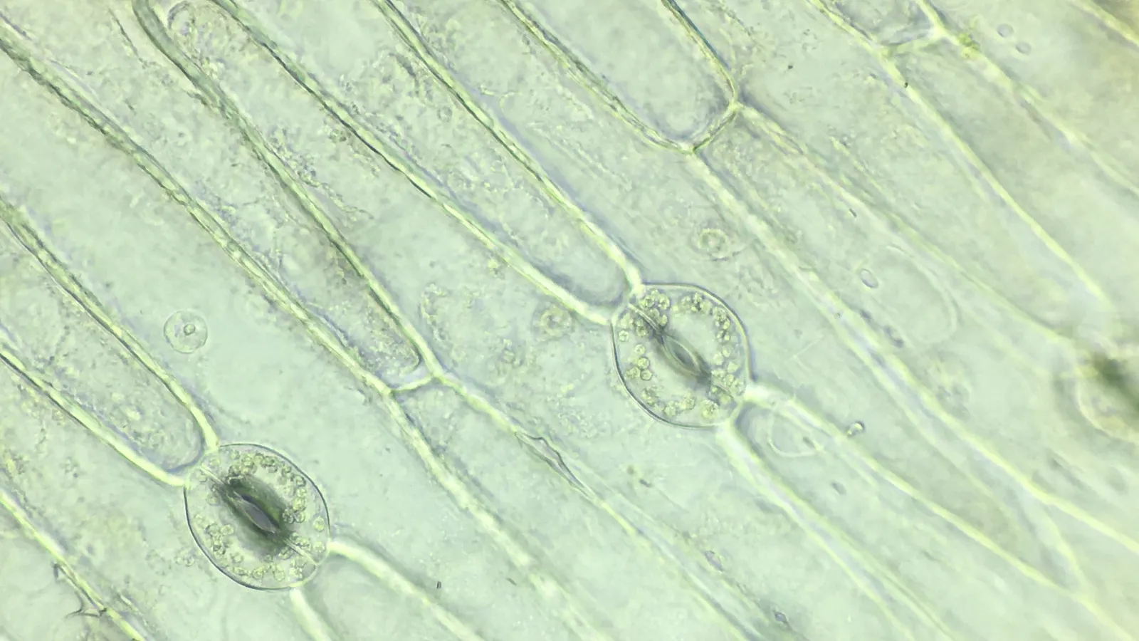
You will see a sheet of epidermal cells, with small pores scattered between them. Each pore is a stoma.
The NCERT lab manual describes a stoma as "an elliptical pore with two kidney shaped guard cells on either side". The guard cells have thin outer walls and thick inner walls. When the guard cells are turgid (swollen with water) the stoma opens, and when they are flaccid it closes. If you repeat the practical with grass, a monocot, the manual's observation table asks whether the guard cells are bean-seed shaped or dumbbell shaped; grass guard cells are the dumbbell type.
Under high power, look for small dots inside each guard cell. Those are chloroplasts. In a stained peel you may also pick out a nucleus in each guard cell.
How should you draw stomata and guard cells?
Draw two or three stomata with a few surrounding epidermal cells, not the whole field. Use this checklist before you hand in your file:
Two kidney-shaped guard cells facing each other, with the pore between them
The inner wall (next to the pore) drawn thicker than the outer wall
A nucleus in each guard cell
Several small chloroplasts in each guard cell
A few epidermal cells around the stoma
Labels: stoma (stomatal pore), guard cell, chloroplast, nucleus, epidermal cell
A title, such as "Stomata in the lower epidermis of a dicot leaf (stained)"
What do examiners ask in the viva?
The NCERT manual ends with questions that make good viva practice. Here is a short script you can rehearse aloud, with answers based on the manual and on CBSE's answer key:
What is the function of guard cells? They open and close the stoma. They swell (turgid) to open it and go limp (flaccid) to close it.
Why are there more stomata on the lower surface? Fewer on the sunlit upper surface means less water lost through transpiration.
What shape are the guard cells in grass? Dumbbell-shaped.
Why stain with safranin? So the cells are easier to see; they show up pinkish red.
If you cannot yet explain why a swollen guard cell opens the pore, the Fermi Concept Library covers every concept in physics, chemistry, biology, mathematics and computing, each with its own lesson, so you can fill the gap before the viva.
Before practical day, rehearse focusing a compound microscope from low to high power, read how lenticels and stomata handle gas exchange, and go through identifying slides and specimens in biology practicals.