On this page
Put a thin purple peel from the underside of a Rhoeo leaf in salt solution and the coloured part of each cell pulls inward while the cell outline stays put. Move it to plain water and the colour fills the cell again. That is plasmolysis and its reverse, and it is on your syllabus: the CBSE Biology curriculum for 2026-27 lists the study of plasmolysis in epidermal peels, such as Rhoeo or lily leaves or the fleshy scale leaves of an onion bulb, as Experiment 4 for Class 11.
What is plasmolysis, in one line?
Plasmolysis is the detachment of a plant cell's living protoplast from its cell wall when the cell loses water in a strong (hyperosmotic) solution, as described in a review by Ingeborg Lang and colleagues in the journal Plants (Lang et al., 2014). The same review says the process "is mainly driven by the vacuole", and that plasmolysis is reversible (deplasmolysis) and characteristic of living plant cells.
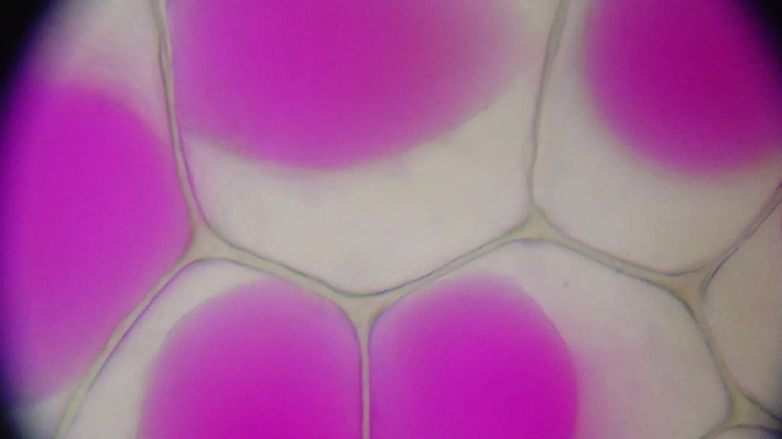
Rhoeo is an older name for Tradescantia spathacea, the oyster plant or Moses-in-the-cradle, whose leaves are dark green above and glossy purple underneath because of anthocyanin pigments (Tradescantia spathacea, Wikipedia). In red onion epidermis the anthocyanin sits in the vacuole (Collings, 2019), and a micrograph of plasmolysed Rhoeo epidermis on Wikimedia Commons shows the coloured vacuoles shrunk inside the cell walls. So in a Rhoeo peel, the purple patch you see is the vacuole, the very part that loses water, and you need no stain.
What shrinks in salt solution, and what does not?
Follow the water:
The salt solution outside has less free water than the cell sap, so water leaves the cell by osmosis (exosmosis).
Most of that water comes out of the vacuole, so the purple area gets smaller.
The cytoplasm and plasma membrane follow the shrinking vacuole and pull away from the wall, often first at the corners, then all round.
The cell wall keeps its shape. It is rigid, so the cell outline does not change.
The space between the wall and the shrunken protoplast fills with the outside solution, because the wall lets it through.
Lang et al. call that gap the periplasmic space. For why plants have a rigid wall at all, see our explainer on how cell walls differ in plants, fungi and bacteria.
To see it clearly, read our guide to focusing a compound microscope step by step and our post on the tonoplast and the plant vacuole.
What happens when you put the peel back in water?
Now the outside water is more dilute than the cell sap, so water moves in (endosmosis), the vacuole swells and the protoplast presses back against the wall. This is deplasmolysis, and since Lang et al. describe plasmolysis as characteristic of living cells, it is your evidence that the cells were alive. If the colour does not come back, the cells may have died; say so in your record instead of copying the expected result.
If hypertonic, hypotonic and water potential still feel like words rather than pictures, the Fermi Concept Library has a lesson for each, so you can connect osmosis to this slide before the viva.
How do you write the plasmolysis observation in your practical record?
In the CBSE Class 11 practical, out of 30 marks, "Practical Record + Viva Voce" carries 4 marks and slide preparation (Experiments 2, 4 and 5, which includes plasmolysis) carries 5 (CBSE Biology curriculum 2026-27). Adapt this template to what you actually saw:
Aim: To study plasmolysis in epidermal peels of a Rhoeo leaf.
Procedure: Mount a piece of the purple lower epidermis in water and observe. Keep a second piece in salt solution for a few minutes, mount it in that solution and observe. Transfer it to water and observe again.
Observation: In water, the purple contents fill each cell up to the wall. In salt solution, the purple contents shrink away from the wall, leaving a colourless space between wall and protoplast; the cell walls keep their shape. After transfer to water, the contents swell and fill the cells again.
Inference: The salt solution was hypertonic to the cell sap, so the cells lost water by exosmosis and were plasmolysed. In water (hypotonic) they gained water by endosmosis and were deplasmolysed. The cells were living.
Precautions: Use a thin, single-layer peel; keep it wet; mount the salt-treated peel in salt solution; avoid air bubbles.
Add two labelled diagrams side by side: a turgid cell (protoplast against the wall) and a plasmolysed cell (cell wall, plasma membrane, shrunken vacuole, and the space filled with outside solution).
To practise the direction-of-water questions that often follow this experiment, the Fermi Problem Library has worked problems with a step-by-step solver that explains the reasoning rather than printing an answer.
Peel, salt, water, then write what you saw before what it means.