On this page
Under a hand lens, a planarian shows two dark eyespots that make it look cross-eyed. They are not camera-style eyes and do not form a detailed picture. They tell the worm which side the light is coming from, and the worm uses that to head for the dark. Here is how, plus a class experiment.
What is a planarian eyespot made of?
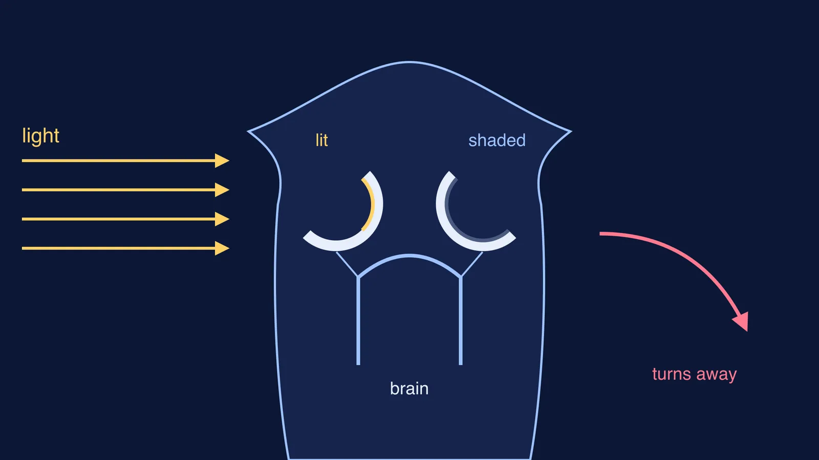
A planarian eye has only two cell types: pigment cells and photoreceptor neurons, according to a 2014 study in PLOS ONE by Paskin and colleagues. The pigment cells form a half-moon-shaped cup that absorbs light. The light-sensing ends of the photoreceptor neurons sit inside the cup.
The cup is open on one side, so light reaches the photoreceptors mainly from the direction the opening faces. The paper notes that each eyecup gives left-right directional information, and the eyes' position at the front of the head adds what is ahead.
Picture light coming from the worm's left. It shines into the left eyecup, but the right eyecup's pigment blocks it. One eye says "bright", the other "dim". That difference is the direction signal.
Do planarians have a brain?
Yes, a simple one. In *Dugesia japonica*, the planarian central nervous system is a mass of cephalic (head) ganglia plus a pair of ventral nerve cords, according to Agata and colleagues in Zoological Science (1998). The ganglia form an inverted U-shaped, brain-like structure with nine branches on each outer side. The two eyes sit on the 3rd branch, and the visual nerve fibres cross to form an optic chiasma inside the U.
The same paper says planarians are considered among the most primitive animals to have developed a central nervous system. Hydra has only a nerve net; our post on how Hydra manages without a brain shows the contrast.
Why do planaria move away from light?
Planarians are photophobic: when exposed to light, they seek cover, as the PLOS ONE paper puts it.
The colour of the light matters. In that study, green, blue and both ultraviolet wavelengths sent at least 80% of worms into the darkest quarter of the test dish by the end of the 2-minute trial. Responses to red and infrared light were not statistically different from controls.
The brain does more than flip a switch. A team at inStem and NCBS in Bengaluru found that a 25 nm change in wavelength is enough to completely switch the worms' light behaviour, even though the eye has a single opsin. The worms compare small differences in how much light each eye absorbs. The same study found that freshly decapitated worms show no significant movement in visible light, but do move away from long-wave UV light, through a reflex-like, whole-body sense that does not need the eyes.
How can you observe light avoidance in class?
This is a short, gentle test, adapted from the four-quarter dish design in the Paskin study. Get planarians from your teacher or a biological supplier, and handle them with a soft paintbrush.
Fill a clear rectangular dish with a shallow layer of the water the worms live in (not tap water straight from the tap).
Draw lines on paper under the dish to split it into four equal quarters, numbered 1 to 4.
Put the dish in a dim room. Gently place 5 or 6 worms in the middle and let them settle.
Shine a phone torch from above one end, over quarter 1. Cover quarter 4 with a card so it is darkest.
Every 30 seconds for 2 minutes, count how many worms are in each quarter.
Run a control: repeat with the torch off.
Swap ends: put the torch over quarter 4 and the card over quarter 1. If the worms follow the dark, it is the light, not the dish, that moves them.
Put the worms back in their container in the dark when you finish.
Record your prediction, the counts per quarter, and one sentence linking the result to the eyecups.
For designing the control, see our guide to control groups in biology experiments. The earthworm light-and-moisture choice experiment uses the same choice-chamber logic with a different animal.
How do you remember this for exams?
Learn it as one chain: the pigment cup shades one side, so the two eyes get unequal light, so the brain turns the worm towards the darker side. Practise drawing the cup.
If ocelli, ganglia and taxis feel like separate facts, the Fermi Concept Library gives each biology concept its own lesson and shows where it sits in the map. And if you want to talk through your experiment results, Fermi's AI tutor can draw the figure and check your understanding as it goes.