On this page
A peptide bond drawn as a plain C–N single line is wrong. The bond sits between a single and a double bond, and that explains why a protein backbone is built from flat, rigid tiles joined at swivels.
Why is the peptide bond planar?
A peptide bond is the amide link, –CO–NH–, joining the carbonyl carbon of one amino acid to the nitrogen of the next. Nitrogen's lone pair sits right next to a C=O group. The OpenStax Organic Chemistry textbook/26:_Biomolecules-_Amino_Acids_Peptides_and_Proteins/26.04:_Peptides_and_Proteins) explains that it is delocalised by resonance with the carbonyl group, which gives the C–N bond a certain amount of double-bond character and restricts rotation around it.
You can draw it as two resonance structures:
Form 1: C=O double bond, C–N single bond, lone pair on N.
Form 2: C–O⁻ single bond, C=N⁺ double bond, no lone pair on N.
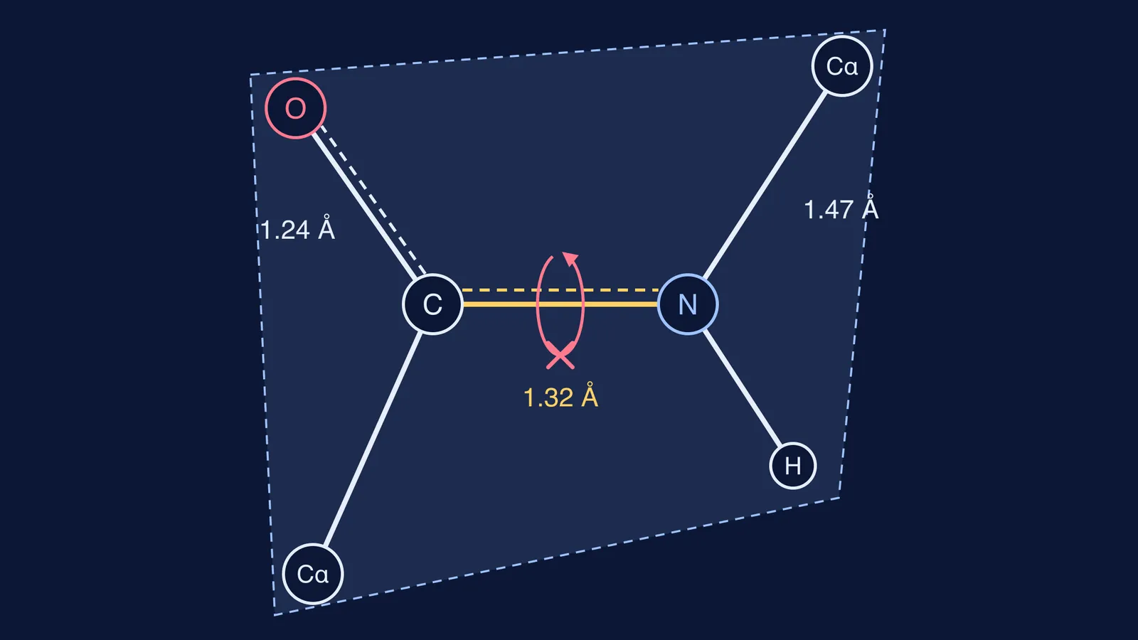
The real bond is a blend. Sharing electrons across C, N and O needs parallel p orbitals, which only happens if the atoms around them lie in one plane. In their 1953 paper on the fundamental dimensions of polypeptide chains, Robert Corey and Linus Pauling wrote that the normal coplanarity of the amide group is the result of resonance, which gives the peptide bond partial double-bond character. Six atoms share that plane: the two α-carbons, the carbonyl C and O, and the N with its H.
How do bond lengths prove partial double-bond character?
Here is a worked check using the values in the Corey and Pauling abstract, drawn from X-ray studies of amide and peptide crystals. C′ is the carbonyl carbon.
Compare the two bonds from the same nitrogen. The peptide bond N–C′ is 1.32 Å. The ordinary N–Cα single bond on the other side is 1.47 Å.
Find the difference: 1.47 − 1.32 = 0.15 Å, so the peptide bond is about 10% shorter. Extra electron density pulls atoms closer, so it has more than single-bond character.
Add the angles round the carbonyl carbon: 125° (N–C′–O) + 114° (N–C′–Cα) + 121° (O–C′–Cα) = 360°. The carbon is flat, as sp² carbon should be.
Do the same round nitrogen: 123° (Cα–N–C′) + 114° (Cα–N–H) + 123° (H–N–C′) = 360°. This nitrogen is flat, not pyramidal like ammonia's, which shows its lone pair is busy in resonance.
Why can't the peptide bond rotate freely?
Turning the C–N bond would twist nitrogen's p orbital away from carbon's and break the π overlap. A LibreTexts biochemistry module on secondary structure says this is why rotation is unfavourable; Corey and Pauling called it severely restricted. Alkenes work the same way, as we explain in why rotation about a carbon-carbon double bond is restricted.
So a peptide bond is locked as trans (α-carbons on opposite sides) or cis. For residues other than proline, the same module says cis is less stable by about 15 kJ/mol, giving a trans to cis ratio of about 1000:1. Proline is the exception: Jakubowski and Flatt's Fundamentals of Biochemistry/01:_Unit_I-_Structure_and_Catalysis/04:_The_Three-Dimensional_Structure_of_Proteins/4.01:_Main_Chain_Conformations) gives a trans to cis ratio of about 4:1 for X–Pro bonds in proteins.
The backbone still bends. The bonds either side of each α-carbon, measured by the angles φ (phi) and ψ (psi), can turn from −180° to +180°, the same textbook notes. A protein chain is flat amide tiles joined at swivels.
Why does a flat peptide bond matter for protein shape?
Planarity was a key assumption behind the α-helix. In a PNAS perspective on the discovery, David Eisenberg describes how Pauling, Corey and Herman Branson, in a paper submitted on 28 February 1951, built helices with planar amide groups and crystal bond dimensions. Only two fitted; the one with 3.7 residues per turn became the α-helix. Earlier models by Bragg, Kendrew and Perutz, Eisenberg notes, had not enforced planar peptide groups. The full story is in how Linus Pauling found the alpha helix.
To trace how resonance in chemistry connects to protein structure in biology, the Fermi Concept Library gives each concept its own lesson and its place in the map.
How do you answer 'why is the peptide bond planar' in an exam?
Use this five-point answer for a 2- or 3-mark board-exam question (numbers from Corey and Pauling):
Draw both resonance structures of –CO–NH–, with curved arrows from N's lone pair and from the C=O π bond.
State that C–N gets partial double-bond character.
Give the evidence: C–N (1.32 Å) is shorter than the N–Cα single bond (1.47 Å).
State the consequence: six atoms lie in one plane and rotation about C–N is restricted.
Add that trans is preferred, and the chain turns about φ and ψ instead.
For drawing practice, see peptide bonds: drawing and naming dipeptides like Gly-Ala. If you get a question wrong and cannot see why, the Fermi Problem Library has worked problems with a step-by-step solver that explains the reasoning rather than printing an answer.