On this page
A biology question gives you a photomicrograph, a scale bar or a reading in eyepiece units, and asks how big the cell really is. One rearranged formula and one unit conversion solve all three.
What is the formula for actual size?
The WJEC exam board's microscope calibration guide gives the relationship as image size = actual size × magnification, or I = A × M. Rearranged, actual size = image size ÷ magnification (A = I ÷ M). The same guide adds one warning: check that the image size and the actual size are in the same units.
You measure the image in millimetres, but cells are measured in micrometres. In the SI system, milli means 10⁻³ and micro means 10⁻⁶, according to the BIPM table of SI prefixes. So 1 mm = 1000 µm. Convert your ruler reading to µm before you divide, and the answer comes out in µm.
Solved question 1. A photomicrograph at ×1500 shows a human red blood cell 10.5 mm across. What is its actual diameter?
Convert: 10.5 mm = 10 500 µm.
Divide: 10 500 µm ÷ 1500 = 7.0 µm.
That matches the NCERT Class 11 chapter Cell: The Unit of Life, which says human red blood cells are about 7.0 µm in diameter.
How do you calculate actual size from a scale bar?
Some questions give a scale bar instead. Use the bar to find the magnification, then use the formula as before.
Measure the scale bar with a ruler, in mm.
Convert that length to µm.
Magnification = measured bar length ÷ the length written on the bar.
Measure the cell in the image and convert to µm.
Actual size = image size ÷ magnification.
Solved question 2. A scale bar labelled 10 µm measures 20 mm on the page. A cell in the same image measures 35 mm.
Bar: 20 mm = 20 000 µm, so magnification = 20 000 ÷ 10 = ×2000.
Cell: 35 mm = 35 000 µm, so actual size = 35 000 ÷ 2000 = 17.5 µm.
How do you use an eyepiece graticule to measure a cell?
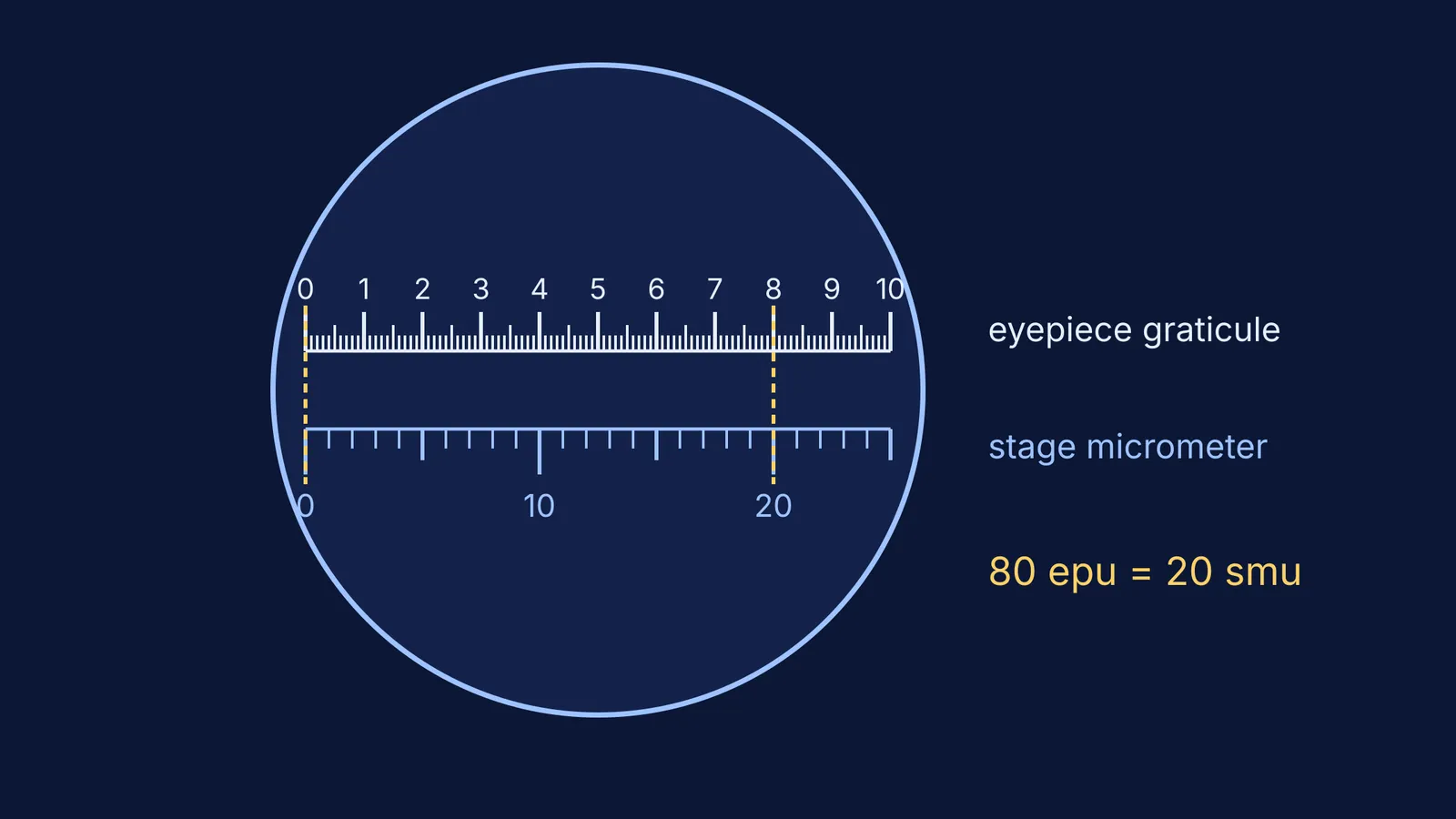
An eyepiece graticule is a scale inside the eyepiece. The WJEC guide describes one marked 1 to 10 with 10 subdivisions between each number, giving 100 eyepiece units (epu). At different magnifications, each division covers a different length of the specimen.
So you calibrate it with a stage micrometer. On the 1 mm type, each of the 100 stage micrometer units (smu) is 0.01 mm, or 10 µm, according to the same guide; a 10 mm type has 0.1 mm units, so check which you have.
Line up the zero of the graticule with the zero of the stage micrometer.
Make sure the two scales are parallel.
Find the next point where the two scales line up again.
Count the divisions on each scale between the two points.
Solved question 3. In the WJEC example, with the ×40 objective, 80 epu line up with 20 smu. The stage micrometer's divisions are 10 µm.
20 smu = 20 × 10 µm = 200 µm.
1 epu = 200 µm ÷ 80 = 2.5 µm.
Swap in your slide without changing the objective. A cell that spans 12 epu is 12 × 2.5 µm = 30 µm long.
Recalibrate every time you change the objective lens. If the scales look blurred, our guide to focusing a compound microscope goes through it step by step.
How do you know your answer is sensible?
Compare it with sizes you already know. The NCERT chapter Cell: The Unit of Life gives these:
Mycoplasmas, the smallest cells, are only 0.3 µm long.
Bacteria can be 3 to 5 µm long.
Human red blood cells are about 7.0 µm in diameter.
Solved question 4. A bacterium measures 6 mm in a photomicrograph at ×1500. 6 mm = 6000 µm, and 6000 ÷ 1500 = 4 µm, inside the 3 to 5 µm range. Skip the conversion and write 0.004 µm, and your bacterium is smaller than any cell.
Before you move on, check:
Both lengths in the same unit, usually µm, before dividing.
Divide for actual size; multiply only when finding image size.
Magnification worked out from the scale bar when one is given.
Graticule recalibrated for the objective you are using.
Answer has a unit and fits what you know about that kind of cell.
For more reference sizes, read the largest and smallest cells, with examples, and for what a light microscope cannot show, see light versus electron microscopes.
If the unit step keeps tripping you up, the Fermi Problem Library has worked problems with a step-by-step solver that explains the reasoning rather than printing an answer, so you can see where a conversion slipped. The Fermi Concept Library covers every concept in physics, chemistry, biology and mathematics, each with its own lesson.
Tomorrow, redo the four questions with the answers covered.