On this page
When a doctor taps just below your kneecap and your foot swings forward, you have not decided anything. The kick is arranged inside your spinal cord, and the loop finishes long before a deliberate movement could start. Here is each step, and how to write it up in an exam.
How does the knee jerk reflex work?
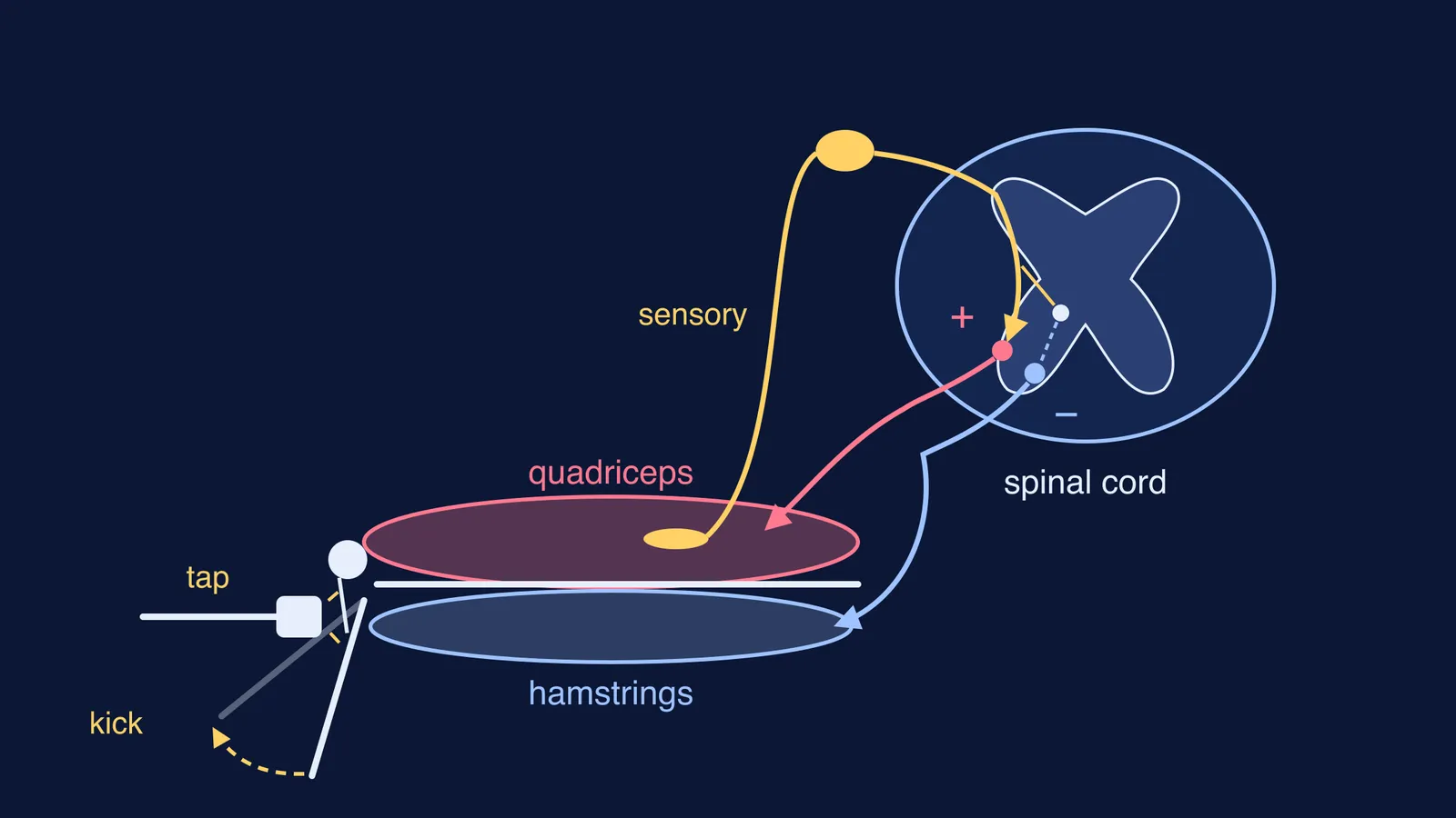
The knee jerk, or patellar reflex, is a stretch reflex: a muscle contracts because it has just been stretched. The loop has five parts, set out in this physiology lab chapter by Karri Haen Whitmer on LibreTexts/02%3A_Labs/2.07%3A_Patellar_and_Achilles_Reflexes):
Stimulus. The hammer strikes the patellar tendon just below the kneecap, which pulls the quadriceps, the big muscle on the front of the thigh, slightly longer.
Receptor. Muscle spindles, small stretch-sensing structures lying alongside the ordinary muscle fibres, detect that sudden stretch.
Sensory neuron. A sensory neuron carries the signal from the spindle into the spinal cord.
One synapse. Inside the cord, the sensory neuron connects directly to a motor neuron, so only one synapse is crossed. That is why the knee jerk is called monosynaptic.
Effector. The motor neuron carries the signal back to the quadriceps, the muscle contracts, and your lower leg kicks forward.
The same chapter notes that the patellar reflex tests the integrity of the L2 to L4 segments of the spinal cord.
If the difference between a sensory neuron, a motor neuron and a synapse still feels blurry, the Fermi Concept Library gives every concept in biology its own lesson and its place in the map, so you can see how the reflex arc connects to them. Our explainer on the structure of a synapse, with a labelled diagram is a good companion too.
Why do the hamstrings relax at the same time?
A kick needs the muscles on the back of the thigh to get out of the way. While the quadriceps is being told to contract, an inhibitory message is sent through an interneuron to the hamstrings, so they relax, according to the LibreTexts chapter/02%3A_Labs/2.07%3A_Patellar_and_Achilles_Reflexes).
Why is the brain not involved in the knee jerk?
The hand-over from sensing neuron to moving neuron happens in the spinal cord, so the signal does not have to travel up to the brain and back first. The LibreTexts chapter puts it this way: processing at the spinal cord means reflex responses "have a relatively short path length and, thus, a quick reaction time."
The numbers show how quick. In a 2025 study in Frontiers in Neurology, Zakia Khatun and colleagues recorded the electrical activity of the quadriceps in 40 healthy people. In the 25 young adults (under 30), the muscle response began on average 17.68 milliseconds after the tendon was struck; in the 15 older adults (mean age 59.8 years) it began at 20.92 milliseconds.
Now compare a choice you make on purpose. In a 2015 study of 120 first-year medical students at Government Medical College, Patiala, pressing a key on seeing a red circle took 247.60 milliseconds on average, and pressing it on hearing a beep took 228.01 milliseconds. The studies measured different things (a muscle signal versus a key press), so this is a rough comparison, but the knee jerk starts more than ten times sooner.
The brain can still turn the reflex up or down. In a 2012 experiment by Steven Passmore and Paul Bruno, 16 adults had their knee jerk tested while trying to pull their interlocked fingers apart (the Jendrassik manoeuvre) or while doing a colour-word puzzle. Pulling the fingers made the reflex bigger and faster; the mental task made no significant difference. The brain does not trigger the kick, but it can change how strongly the cord responds.
How do you write the knee jerk reflex arc in an exam?
Use this order, and draw the arrows in the same order:
Name it: a monosynaptic stretch reflex, with the spinal cord as the centre.
Stimulus: a tap on the patellar tendon stretches the quadriceps.
Receptor: muscle spindles in the quadriceps sense the stretch.
Sensory neuron carries the impulse into the spinal cord.
Synapse: the sensory neuron connects directly with a motor neuron.
Motor neuron carries the impulse back; the quadriceps contracts and the leg kicks.
Add: an interneuron inhibits the hamstrings so they relax.
In your diagram, check that arrows run from receptor to muscle, the receptor sits in the thigh muscle, not the knee, and no line goes to the brain.
Next, read our guide to voluntary, involuntary and reflex actions, and for the history, our profile of Charles Sherrington and his work on reflexes and the synapse.