On this page
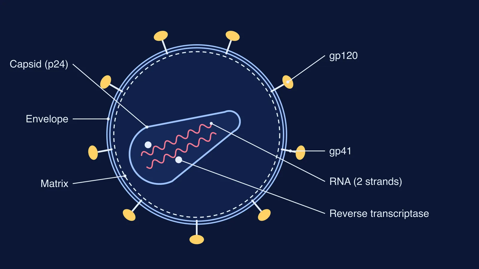
If a test asks for a labelled diagram of HIV, a circle with spikes and a squiggle inside leaves out most of what makes HIV recognisable. A good diagram puts the parts in the right places: spikes that cross the envelope, a cone-shaped core, and two separate RNA strands with an enzyme beside them. This guide gives you seven labels, the order to draw them in, and a checklist for the mistakes that cost marks.
What are the parts of HIV, from outside to inside?
HIV is built in layers. The labels below follow the review HIV-1 Assembly, Budding, and Maturation by Wesley Sundquist and Hans-Georg Kräusslich (Cold Spring Harbor Perspectives in Medicine, 2012), unless another source is linked.
Envelope. A lipid bilayer taken from the host cell: the virus “acquires its lipid envelope and Env protein spikes as it buds from the plasma membrane”, the review says.
gp120. The surface glycoprotein that forms the “head” of each spike, as described in a 2006 Nature study of AIDS virus envelope spikes.
gp41. The transmembrane glycoprotein that forms the stalk and anchors the spike in the envelope. Each spike is a trimer: three gp120 heads on three gp41 stalks.
Matrix. Matrix protein (MA) stays on the inner face of the envelope as a discontinuous shell.
Capsid. The cone-shaped core. Its shell is made of approximately 1,200 copies of capsid protein (CA), the protein also called p24.
Two RNA strands. HIV packages two copies of its single-stranded, positive-sense RNA genome, bound by nucleocapsid protein (NC).
Reverse transcriptase. One of three viral enzymes packed inside, with protease and integrase.
How big is HIV and how many spikes does it have?
Retroviruses are generally spherical particles 100 to 200 nm across, according to a 2015 review of the morphology of retrovirus particles in AIMS Biophysics. The same review describes the HIV-1 gp120–gp41 spike as mushroom-shaped and about 120 Å (12 nm) tall.
In the 2006 Nature study, Ping Zhu and colleagues used cryo-electron tomography and found about 14 spikes per wild-type HIV-1 particle, with some spikes clustered together. On an answer sheet, 8 to 10 spikes around the outline keeps the diagram readable.
How do you draw the HIV diagram step by step?
Use a pencil and leave the right third of the space for labels.
Draw a large circle with a second circle just inside it. This double line is the envelope.
Add 8 to 10 spikes: a short stick through the envelope (gp41) topped with a knob (gp120).
Just inside the envelope, draw a dashed circle for the matrix.
In the centre, draw a cone with one wide rounded end and one narrow rounded end. This is the capsid. Do not draw it as a circle.
Inside the cone, draw two separate wavy lines for the two RNA strands.
Add two small dots beside the RNA and label one “reverse transcriptase”.
Draw label lines with a ruler, keep them from crossing, and write the labels in one column.
If you find it easier to learn a figure by watching it built up, Fermi's AI tutor can draw the figure on a board and check understanding as it goes, so you can practise naming each part before the exam.
What does each labelled part do?
A diagram question often ends with “state one function”. One line per part is enough:
Envelope: the outer covering, taken from the host cell membrane as the virus buds out.
gp120 and gp41: Env spikes “initiate infection of host cells”, the Zhu study notes, which is why they are targets for vaccine development.
Matrix: lines the inside of the envelope.
Capsid (p24): helps maintain the structural integrity of the virus and encloses the RNA and enzymes.
RNA: carries the virus's genes.
Reverse transcriptase: an enzyme that uses RNA as a template to make a complementary DNA molecule, which HIV needs to copy its genome.
To follow what happens after the virus enters a cell, read our explainer on how HIV replicates through reverse transcription and helper T cell loss. The story of how the enzyme was found is in how Temin and Baltimore discovered reverse transcriptase.
Which mistakes cost marks on an HIV diagram?
Run this checklist before you move to the next question:
The capsid is a cone, not a circle or a hexagon.
There are two RNA strands, labelled as single-stranded RNA.
Spike stalks pass through the envelope instead of floating outside it.
There is no “cell wall”, “nucleus” or “cytoplasm” label: a virus is not a cell.
At least one enzyme is labelled.
Label lines are straight and do not cross.
Viral diagrams often come in pairs, so practise this one next to the bacteriophage T4 diagram with its head, tail and fibres. If the parts still feel like a list of words, the Fermi Concept Library gives every biology concept its own lesson and its place in the map, so you can see where virus structure fits.
Draw it once today from memory, and check it against the seven labels.