On this page
Your blood carries everything it needs to make a clot, all the time. NCERT's Class 11 chapter on body fluids and circulation notes that the factors for coagulation are present in the plasma in an inactive form. So the real question is not how blood clots, but why it waits. The answer is that clotting is held back everywhere, and released only at a wound.
Why does blood not clot inside blood vessels?
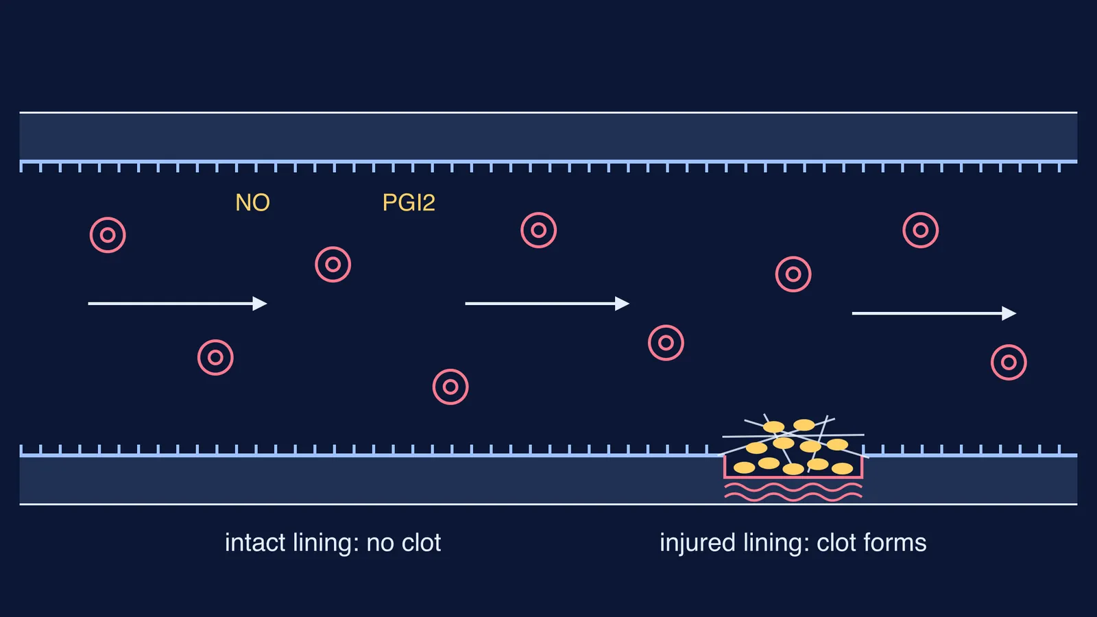
The inside of every vessel is lined by a single layer of cells called the endothelium. A 2021 review, Endothelial cells and coagulation by Neubauer and Zieger in Cell and Tissue Research, describes it as a nonadhesive surface that keeps blood fluid in several ways at once:
Nitric oxide (NO) diffuses into nearby platelets and damps down their activation.
Prostacyclin (PGI2) works independently of NO and also stops platelets from switching on.
Heparan sulfate in the sugary coat on the cell surface (the glycocalyx) binds antithrombin and boosts its activity, so it can inactivate clotting enzymes.
Thrombomodulin binds thrombin and activates protein C, which breaks down clotting factors Va and VIIIa.
Tissue factor pathway inhibitor (TFPI) limits the action of tissue factor, the trigger at the top of the clotting cascade.
Tissue plasminogen activator (t-PA) helps break down any fibrin that does form.
The surface matters too. Platelets need something to grip, and an intact lining keeps collagen and tissue factor hidden underneath. So the lining hides the trigger and keeps brakes on at every level.
What does heparin do, and where does it come from?
In the NCERT chapter, basophils secrete histamine, serotonin and heparin, and basophils are the least common white blood cells, at 0.5 to 1 per cent of the total. Heparin is a close chemical cousin of the heparan sulfate on the vessel wall, and it works the same way: through antithrombin.
According to the American College of Chest Physicians guideline on parenteral anticoagulants (Garcia and colleagues, Chest, 2012), heparin binds antithrombin through a unique pentasaccharide sequence and changes its shape, so antithrombin becomes a rapid inhibitor of thrombin and factor Xa. The same paper notes that only about one-third of heparin molecules carry that pentasaccharide, and that chains shorter than 18 sugar units cannot bridge antithrombin to thrombin, though they can still help it inhibit factor Xa.
The key idea for an exam: heparin does not dissolve clots. It speeds up a natural inhibitor, antithrombin, so new thrombin is mopped up before it can turn fibrinogen into fibrin. Our post on the leech and hirudin shows another animal's way of blocking the same enzyme.
What changes when a vessel is injured?
Injury tears the lining and exposes what lies beneath. The Neubauer and Zieger review explains that exposed subendothelial collagen lets platelets stick, with von Willebrand factor acting as a bridge, while tissue factor, hidden in the intact wall, is exposed to blood and sets off the clotting cascade.
In NCERT's simpler account, an injury or trauma stimulates platelets to release factors that activate coagulation, and factors released by the tissues at the site of injury can also start it. A cascade of reactions forms thrombokinase, which helps convert prothrombin to thrombin; thrombin converts fibrinogen to fibrin threads; and calcium ions play a very important role. Blood normally contains 1,50,000 to 3,50,000 platelets per cubic millimetre, according to the same NCERT chapter. For each step in order, read our walk-through of platelets, thrombin and fibrin in the clotting cascade.
How do you write this answer in a biology exam?
Use this four-step frame for a 3-mark or 5-mark answer.
State the fact. Clotting factors circulate in inactive form, so clotting needs a trigger.
Name the barrier. The intact endothelium is nonadhesive and hides collagen and tissue factor.
Name the brakes. Heparin (from basophils) and heparan sulfate work with antithrombin; the lining also releases NO and prostacyclin, which keep platelets inactive.
Contrast with injury. Damage exposes collagen and tissue factor, platelets stick, and the cascade runs: prothrombin to thrombin, fibrinogen to fibrin, with Ca2+.
Check your answer against these common slips:
Writing that heparin "breaks down clots". It prevents new fibrin forming; it does not dissolve old clots.
Saying platelets are absent from vessels. They circulate all the time, just not activated.
Forgetting calcium. Many questions test it.
Mixing up fibrinogen (soluble, in plasma) with fibrin (the threads).
If the cell names blur, revise the formed elements of blood: RBCs, WBC types and platelets first. The Fermi Concept Library covers every concept in biology, each with its own lesson and its place in the map. For practice questions, the Fermi Problem Library has worked problems with a step-by-step solver that explains the reasoning rather than printing an answer.
Next time you revise this chapter, try explaining it aloud in four sentences without notes.